HRP

HRP

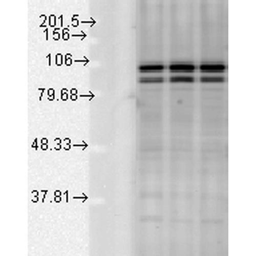
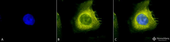
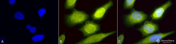
  • Immunocytochemistry/Immunofluorescence analysis using Rabbit Anti-AHA1 Polyclonal Antibody (STPA00032). Tissue: Heat Shocked Cervical cancer cell line (HeLa). Species: Human. Fixation: 2% Formaldehyde for 20 min at RT. Primary Antibody: Rabbit Anti-AHA1 Polyclonal Antibody (STPA00032) at 1:60 for 12 hours at 4°C. Secondary Antibody: R-PE Goat Anti-Rabbit (yellow) at 1:200 for 2 hours at RT. Western blot analysis of multiple Cell line lysates showing detection of AHA1 protein using Rabbit Anti-AHA1 Polyclonal Antibody (STPA00032). Load: 15 µgprotein. Block: 1.5% BSA for 30 minutes at RT. Primary Antibody: Rabbit Anti-AHA1 Polyclonal Antibody (STPA00032) at 1:1000 for 2 hours at RT. Secondary Antibody: Donkey Anti-Rabbit IgG: HRP for 1 hour at RT.

    Anti-AHA1 Antibody

    AHA1 AntibodyAHA1 Antibody is a rabbit polyclonal antibody directed against AHA1, offered by Assay Genie for research applications. Reported applications include WB, IHC, ICC/IF, IP and ELISA...
  • Immunocytochemistry/Immunofluorescence analysis using Rabbit Anti-Calnexin-CT Polyclonal Antibody (STPA00031). Tissue: Heat Shocked Cervical cancer cell line (HeLa). Species: Human. Fixation: 2% Formaldehyde for 20 min at RT. Primary Antibody: Rabbit Anti-Calnexin-CT Polyclonal Antibody (STPA00031) at 1:80 for 12 hours at 4°C. Secondary Antibody: FITC Goat Anti-Rabbit (green) at 1:200 for 2 hours at RT. Western blot analysis of Rat tissue mix showing detection of Calnexin-CT protein using Rabbit Anti-Calnexin-CT Polyclonal Antibody (STPA00031). Load: 15 µgprotein. Block: 1.5% BSA for 30 minutes at RT. Primary Antibody: Rabbit Anti-Calnexin-CT Polyclonal Antibody (STPA00031) at 1:1000 for 2 hours at RT. Secondary Antibody: Donkey Anti-Rabbit IgG: HRP for 1 hour at RT.

    Anti-Calnexin-CT Antibody

    Calnexin-CT AntibodyCalnexin-CT Antibody is a rabbit polyclonal antibody directed against Calnexin-CT, offered by Assay Genie for research applications. Reported applications include WB, IHC, ICC/IF,...
  • Immunocytochemistry/Immunofluorescence analysis using Rabbit Anti-GRP78 (Bip) Polyclonal Antibody (STPA00029). Tissue: Heat Shocked Cervical cancer cell line (HeLa). Species: Human. Fixation: 2% Formaldehyde for 20 min at RT. Primary Antibody: Rabbit Anti-GRP78 (Bip) Polyclonal Antibody (STPA00029) at 1:100 for 12 hours at 4°C. Secondary Antibody: FITC Goat Anti-Rabbit (green) at 1:200 for 2 hours at RT. Western blot analysis of Human Glucose deprived glia cell lysates showing detection of GRP78 protein using Rabbit Anti-GRP78 Polyclonal Antibody (STPA00029). Primary Antibody: Rabbit Anti-GRP78 Polyclonal Antibody (STPA00029) at 1:1000.

    Anti-GRP78 (Bip) Antibody

    GRP78 (Bip) AntibodyGRP78 (Bip) Antibody is a rabbit polyclonal antibody directed against GRP78 (Bip), offered by Assay Genie for research applications. Reported applications include WB, IHC, ICC/IF...
  • Immunocytochemistry/Immunofluorescence analysis using Rabbit Anti-HSP22 Polyclonal Antibody (STPA00030). Tissue: HaCaT cells. Species: Human. Fixation: Cold 100% methanol at -20C for 10 minutes. Primary Antibody: Rabbit Anti-HSP22 Polyclonal Antibody (STPA00030) at 1:100 for 12 hours at 4°C. Secondary Antibody: FITC Goat Anti-Rabbit at 1:50 for 1-2 hours at RT in dark. Localization: Nuclear Staining. Western blot analysis of Rat Skeletal muscle lysates showing detection of HSP22 protein using Rabbit Anti-HSP22 Polyclonal Antibody (STPA00030). Load: 15 µgprotein. Block: 1.5% BSA for 30 minutes at RT. Primary Antibody: Rabbit Anti-HSP22 Polyclonal Antibody (STPA00030) at 1:1000 for 2 hours at RT. Secondary Antibody: Donkey Anti-Rabbit IgG: HRP for 1 hour at RT.

    Anti-HSP22 Antibody

    HSP22 AntibodyHSP22 Antibody is a rabbit polyclonal antibody directed against HSP22, offered by Assay Genie for research applications. Reported applications include WB, IHC, ICC/IF and IP. Reported...
  • Immunocytochemistry/Immunofluorescence analysis using Chicken Anti-Hsp70 Polyclonal Antibody (STPA00028). Tissue: Heat Shocked Cervical cancer cell line (HeLa). Species: Human. Fixation: 2% Formaldehyde for 20 min at RT. Primary Antibody: Chicken Anti-Hsp70 Polyclonal Antibody (STPA00028) at 1:120 for 12 hours at 4°C. Secondary Antibody: R-PE Goat Anti-Chicken (yellow) at 1:200 for 2 hours at RT. Western blot analysis of Human Cervical cancer cell line (HeLa) lysate showing detection of HSP70 protein using Chicken Anti-HSP70 Polyclonal Antibody (STPA00028). Load: 15 µgprotein. Block: 1.5% BSA for 30 minutes at RT. Primary Antibody: Chicken Anti-HSP70 Polyclonal Antibody (STPA00028) at 1:1000 for 2 hours at RT. Secondary Antibody: Goat Anti-Chicken IgY: HRP for 1 hour at RT.

    Anti-HSP70 Antibody

    HSP70 AntibodyHSP70 Antibody is a chicken polyclonal antibody directed against HSP70, offered by Assay Genie for research applications. Reported applications include WB, ICC/IF and ELISA. Reported...
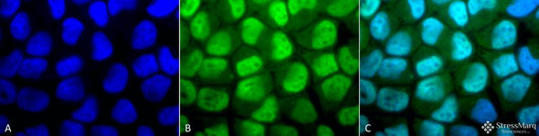
  • Immunocytochemistry/Immunofluorescence analysis using Rabbit Anti-p38 Polyclonal Antibody (STPA00027). Tissue: Cervical cancer cell line (HeLa). Species: Human. Fixation: 2% Formaldehyde for 20 min at RT. Primary Antibody: Rabbit Anti-p38 Polyclonal Antibody (STPA00027) at 1:100 for 12 hours at 4°C. Secondary Antibody: FITC Goat Anti-Rabbit (green) at 1:200 for 2 hours at RT. Counterstain: DAPI (blue) nuclear stain at 1:40000 for 2 hours at RT. Western blot analysis of Human cancer cell lines showing detection of p38 protein using Rabbit Anti-p38 Polyclonal Antibody (STPA00027). Load: 15 µgprotein. Block: 1.5% BSA for 30 minutes at RT. Primary Antibody: Rabbit Anti-p38 Polyclonal Antibody (STPA00027) at 1:4000 for 2 hours at RT. Secondary Antibody: Donkey Anti-Rabbit IgG: HRP for 1 hour at RT.

    Anti-p38 Antibody

    p38 Antibodyp38 Antibody is a rabbit polyclonal antibody directed against p38, offered by Assay Genie for research applications. Reported applications include WB, IHC, ICC/IF and IP. Reported...
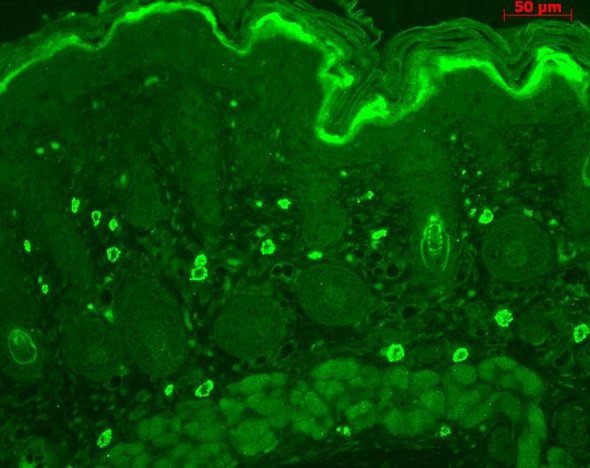
  • Immunocytochemistry/Immunofluorescence analysis using Rabbit Anti-TNF-R1 Polyclonal Antibody (STPA00026). Tissue: Cervical cancer cell line (HeLa). Species: Human. Fixation: 2% Formaldehyde for 20 min at RT. Primary Antibody: Rabbit Anti-TNF-R1 Polyclonal Antibody (STPA00026) at 1:100 for 12 hours at 4°C. Secondary Antibody: APC Goat Anti-Rabbit (red) at 1:200 for 2 hours at RT. Immunohistochemistry analysis using Rabbit Anti-TNF-R1 Polyclonal Antibody (STPA00026). Tissue: backskin. Species: Mouse. Fixation: Bouin's Fixative Solution. Primary Antibody: Rabbit Anti-TNF-R1 Polyclonal Antibody (STPA00026) at 1:100 for 1 hour at RT. Secondary Antibody: FITC Goat Anti-Rabbit (green) at 1:50 for 1 hour at RT. Localization: dermis.

    Anti-TNF-R1 Antibody

    TNF-R1 AntibodyTNF-R1 Antibody is a rabbit polyclonal antibody directed against TNF-R1, offered by Assay Genie for research applications. Reported applications include WB, IHC, ICC/IF and IP...
  • Immunocytochemistry/Immunofluorescence analysis using Rabbit Anti-Rab5 Polyclonal Antibody (STPA00025). Tissue: Cervical cancer cell line (HeLa). Species: Human. Fixation: 2% Formaldehyde for 20 min at RT. Primary Antibody: Rabbit Anti-Rab5 Polyclonal Antibody (STPA00025) at 1:80 for 12 hours at 4°C. Secondary Antibody: R-PE Goat Anti-Rabbit (yellow) at 1:200 for 2 hours at RT. Western blot analysis of Human Cell line lysates showing detection of Rab5 protein using Rabbit Anti-Rab5 Polyclonal Antibody (STPA00025). Load: 15 µgprotein. Block: 1.5% BSA for 30 minutes at RT. Primary Antibody: Rabbit Anti-Rab5 Polyclonal Antibody (STPA00025) at 1:1000 for 2 hours at RT. Secondary Antibody: Donkey Anti-Rabbit IgG: HRP for 1 hour at RT.

    Anti-Rab5 Antibody

    Rab5 AntibodyRab5 Antibody is a rabbit polyclonal antibody directed against Rab5, offered by Assay Genie for research applications. Reported applications include WB, IHC and ICC/IF. Reported...
  • Immunocytochemistry/Immunofluorescence analysis using Rabbit Anti-Methylated Lysine Polyclonal Antibody (STPA00024). Tissue: Cervical cancer cell line (HeLa). Species: Human. Fixation: 2% Formaldehyde for 20 min at RT. Primary Antibody: Rabbit Anti-Methylated Lysine Polyclonal Antibody (STPA00024) at 1:50 for 12 hours at 4°C. Secondary Antibody: FITC Goat Anti-Rabbit (green) at 1:200 for 2 hours at RT. Western blot analysis of Bovine serum albumin showing detection of Methylated Lysine protein using Rabbit Anti-Methylated Lysine Polyclonal Antibody (STPA00024). Primary Antibody: Rabbit Anti-Methylated Lysine Polyclonal Antibody (STPA00024) at 1:1000. Methylated Lysine in BSA (Left) and Methylated BSA (Right).

    Anti-Methylated Lysine Antibody

    Methylated Lysine AntibodyMethylated Lysine Antibody is a rabbit polyclonal antibody directed against Methylated Lysine, offered by Assay Genie for research applications. Reported applications...
  • Immunocytochemistry/Immunofluorescence analysis using Rabbit Anti-Acetylated Lysine Polyclonal Antibody (STPA00023). Tissue: Heat Shocked Cervical cancer cell line (HeLa). Species: Human. Fixation: 2% Formaldehyde for 20 min at RT. Primary Antibody: Rabbit Anti-Acetylated Lysine Polyclonal Antibody (STPA00023) at 1:100 for 12 hours at 4°C. Secondary Antibody: R-PE Goat Anti-Rabbit (yellow) at 1:200 for 2 hours at RT. Western blot analysis of Bovine serum albumin showing detection of Acetylated Lysine protein using Rabbit Anti-Acetylated Lysine Polyclonal Antibody (STPA00023). Primary Antibody: Rabbit Anti-Acetylated Lysine Polyclonal Antibody (STPA00023) at 1:1000. Acetylated lysine in BSA (Left) and Acetylated BSA (Right).

    Anti-Acetylated Lysine Antibody

    Acetylated Lysine AntibodyAcetylated Lysine Antibody is a rabbit polyclonal antibody directed against Acetylated Lysine, offered by Assay Genie for research applications. Reported applications...
  • Immunocytochemistry/Immunofluorescence analysis using Rabbit Anti-CDC37 Polyclonal Antibody (STPA00020). Tissue: Heat Shocked Cervical cancer cell line (HeLa). Species: Human. Fixation: 2% Formaldehyde for 20 min at RT. Primary Antibody: Rabbit Anti-CDC37 Polyclonal Antibody (STPA00020) at 1:200 for 12 hours at 4°C. Secondary Antibody: R-PE Goat Anti-Rabbit (yellow) at 1:200 for 2 hours at RT. Western blot analysis of Human Cervical cancer cell line (HeLa) lysate showing detection of CDC37 protein using Rabbit Anti-CDC37 Polyclonal Antibody (STPA00020). Primary Antibody: Rabbit Anti-CDC37 Polyclonal Antibody (STPA00020) at 1:2000.

    Anti-CDC37 Antibody

    CDC37 AntibodyCDC37 Antibody is a rabbit polyclonal antibody directed against CDC37, offered by Assay Genie for research applications. Reported applications include WB and ICC/IF. Reported...
  • Immunocytochemistry/Immunofluorescence analysis using Rabbit Anti-Rab4 Polyclonal Antibody (STPA00019). Tissue: Heat Shocked Cervical cancer cell line (HeLa). Species: Human. Fixation: 2% Formaldehyde for 20 min at RT. Primary Antibody: Rabbit Anti-Rab4 Polyclonal Antibody (STPA00019) at 1:150 for 12 hours at 4°C. Secondary Antibody: R-PE Goat Anti-Rabbit (yellow) at 1:200 for 2 hours at RT. Western blot analysis of Human Cervical cancer cell line (HeLa) lysate showing detection of Rab4 protein using Rabbit Anti-Rab4 Polyclonal Antibody (STPA00019). Load: 15 µgprotein. Block: 1.5% BSA for 30 minutes at RT. Primary Antibody: Rabbit Anti-Rab4 Polyclonal Antibody (STPA00019) at 1:1000 for 2 hours at RT. Secondary Antibody: Donkey Anti-Rabbit IgG: HRP for 1 hour at RT.

    Anti-Rab4 Antibody

    Rab4 AntibodyRab4 Antibody is a rabbit polyclonal antibody directed against Rab4, offered by Assay Genie for research applications. Reported applications include WB, IHC and ICC/IF. Reported...