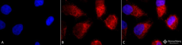
TNF-R1 Antibody is a rabbit polyclonal antibody directed against TNF-R1, offered by Assay Genie for research applications. Reported applications include WB, IHC, ICC/IF and IP. Reported reactivity: Human, Mouse, Rat, Bovine, Monkey, Dog and Rabbit. Purified by Peptide Affinity.
Tumor necrosis factor receptor 1 (TNF-R1), also known as CD120a or p55/p60, is a high-affinity receptor for tumor necrosis factor-alpha (TNF-α) and a key member of the TNF/TNFR superfamily. Structurally, TNF-R1 features an extracellular domain with multiple cysteine-rich repeats and a cytoplasmic death domain that initiates downstream signaling cascades. TNF-R1 is widely expressed in the central nervous system, including neurons, astrocytes, and microglia. Upon binding TNF-α, TNF-R1 activates multiple intracellular pathways, including NF-κB-mediated inflammation and caspase-dependent apoptosis. This dual functionality allows TNF-R1 to regulate both cell survival and programmed cell death, depending on the cellular context and signaling environment. In neurodegenerative disease research, TNF-R1 has emerged as a critical mediator of chronic neuroinflammation and neuronal loss. Aberrant TNF-R1 signaling is implicated in the pathogenesis of Alzheimer’s disease, Parkinson’s disease, multiple sclerosis, and amyotrophic lateral sclerosis (ALS). Sustained activation of TNF-R1 can exacerbate oxidative stress, synaptic dysfunction, and glial reactivity—hallmarks of progressive neurodegeneration. Given its central role in immune signaling and apoptosis, TNF-R1 is a promising therapeutic target for modulating neuroinflammatory responses and protecting neuronal integrity in neurodegenerative disorders. This antibody is also available conjugated to ATTO 390, ATTO 488, ATTO 594, APC, Biotin, FITC, HRP, PerCP and RPE. Supplied in 100 µg. For research use only; not for diagnostic or therapeutic procedures.
1. Kontermann R.E., et al. (2008) J Immunother. 31(3):225-34. 2. Hehlgans T. and Pfeffer K. (2005) Immunology. 115(1):1-20. 3. Al-Lamki S., et al. (2005) The Faseb Journal. 19:1638-1645.



![Anti-TNF alpha [7A8] Monoclonal Antibody, capture (AGMB05977)](https://cdn11.bigcommerce.com/s-rd6ounxcu2/images/stencil/590x590/products/117919/131955/anti-tnf-alpha-7a8-monoclonal-antibody-capture-agmb05977__06328__70328.1786851382.jpg?c=1 "Anti-TNF alpha [7A8] Monoclonal Antibody, capture (AGMB05977)")

![Anti-TNF alpha [5F11] Monoclonal Antibody, unconjugated, detector (AGMB05978)](https://cdn11.bigcommerce.com/s-rd6ounxcu2/images/stencil/590x590/products/117920/131956/anti-tnf-alpha-5f11-monoclonal-antibody-unconjugated-detector-agmb05978__53771__15056.1786851387.jpg?c=1 "Anti-TNF alpha [5F11] Monoclonal Antibody, unconjugated, detector (AGMB05978)")
