HRP

HRP

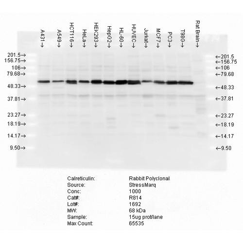
  • Immunocytochemistry/Immunofluorescence analysis using Rabbit Anti-Calreticulin Polyclonal Antibody (STPA00017). Tissue: Heat Shocked Cervical cancer cell line (HeLa). Species: Human. Fixation: 2% Formaldehyde for 20 min at RT. Primary Antibody: Rabbit Anti-Calreticulin Polyclonal Antibody (STPA00017) at 1:100 for 12 hours at 4°C. Secondary Antibody: FITC Goat Anti-Rabbit (green) at 1:200 for 2 hours at RT. Western blot analysis of multiple cell lines lysates showing detection of Calreticulin protein using Rabbit Anti-Calreticulin Polyclonal Antibody (STPA00017). Load: 15 µgprotein. Block: 1.5% BSA for 30 minutes at RT. Primary Antibody: Rabbit Anti-Calreticulin Polyclonal Antibody (STPA00017) at 1:5000 for 2 hours at RT. Secondary Antibody: Donkey Anti-Rabbit IgG: HRP for 1 hour at RT.

    Anti-Calreticulin Antibody

    Calreticulin AntibodyCalreticulin Antibody is a rabbit polyclonal antibody directed against Calreticulin, offered by Assay Genie for research applications. Reported applications include WB, IHC,...
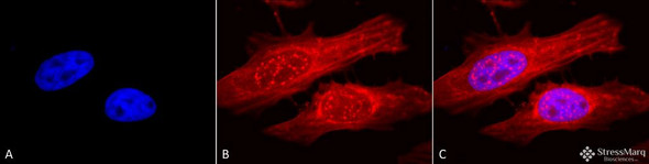
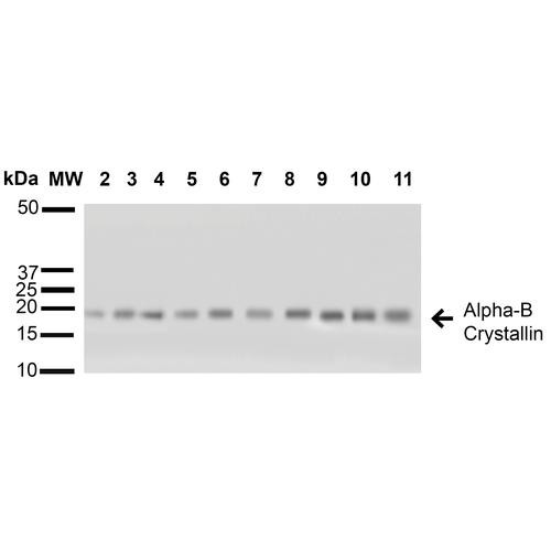
  • Immunocytochemistry/Immunofluorescence analysis using Rabbit Anti-Alpha B Crystallin Polyclonal Antibody (STPA00018). Tissue: Heat Shocked Cervical cancer cell line (HeLa). Species: Human. Fixation: 2% Formaldehyde for 20 min at RT. Primary Antibody: Rabbit Anti-Alpha B Crystallin Polyclonal Antibody (STPA00018) at 1:120 for 12 hours at 4°C. Secondary Antibody: APC Goat Anti-Rabbit (red) at 1:200 for 2 hours at RT. Western blot analysis of Human A431, HCT116, HeLa, HepG2, HEK293, HUVEC, Jurkat, MCF7, PC3 and T98G cell lysates showing detection of ~22 kDa Alpha B Crystallin protein using Rabbit Anti-Alpha B Crystallin Polyclonal Antibody (STPA00018). Lane 1: Molecular Weight Ladder (MW). Lane 2: A431 cell lysates. Lane 3: HCT116 cell lysates. Lane 4: HeLa cell lysates. Lane 5: HepG2 cell lysates. Lane 6: HEK293 cell lysates. Lane 7: HUVEC cell lysates.

    Anti-Alpha B Crystallin Antibody

    Alpha B Crystallin AntibodyAlpha B Crystallin Antibody is a rabbit polyclonal antibody directed against Alpha B Crystallin, offered by Assay Genie for research applications. Reported applications...
  • Immunocytochemistry/Immunofluorescence analysis using Rabbit Anti-Erk1/2 Polyclonal Antibody (STPA00016). Tissue: Cervical cancer cell line (HeLa). Species: Human. Fixation: 2% Formaldehyde for 20 min at RT. Primary Antibody: Rabbit Anti-Erk1/2 Polyclonal Antibody (STPA00016) at 1:100 for 12 hours at 4°C. Secondary Antibody: APC Goat Anti-Rabbit (red) at 1:200 for 2 hours at RT. Western blot analysis of Human Cell line lysates showing detection of ERK1 protein using Rabbit Anti-ERK1 Polyclonal Antibody (STPA00016). Load: 15 µgprotein. Block: 1.5% BSA for 30 minutes at RT. Primary Antibody: Rabbit Anti-ERK1 Polyclonal Antibody (STPA00016) at 1:1000 for 2 hours at RT. Secondary Antibody: Donkey Anti-Rabbit IgG: HRP for 1 hour at RT.

    Anti-Erk1/2 Antibody

    Erk1/2 AntibodyErk1/2 Antibody is a rabbit polyclonal antibody directed against Erk1/2, offered by Assay Genie for research applications. Reported applications include WB, IHC, ICC/IF and FCM...
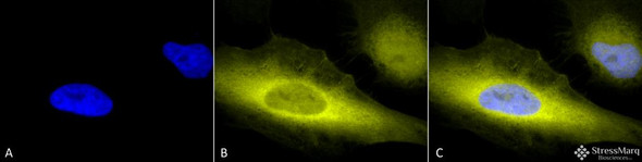
  • Immunocytochemistry/Immunofluorescence analysis using Rabbit Anti-Ubiquitin Polyclonal Antibody (STPA00015). Tissue: Heat Shocked Cervical cancer cell line (HeLa). Species: Human. Fixation: 2% Formaldehyde for 20 min at RT. Primary Antibody: Rabbit Anti-Ubiquitin Polyclonal Antibody (STPA00015) at 1:100 for 12 hours at 4°C. Secondary Antibody: R-PE Goat Anti-Rabbit (yellow) at 1:200 for 2 hours at RT. Western blot analysis of Human Embryonic kidney epithelial cell line (HEK293) lysate showing detection of Ubiquitin protein using Rabbit Anti-Ubiquitin Polyclonal Antibody (STPA00015). Primary Antibody: Rabbit Anti-Ubiquitin Polyclonal Antibody (STPA00015) at 1:1000.

    Anti-Ubiquitin Antibody

    Ubiquitin AntibodyUbiquitin Antibody is a rabbit polyclonal antibody directed against Ubiquitin, offered by Assay Genie for research applications. Reported applications include WB, IHC, ICC/IF and IP...
  • Immunocytochemistry/Immunofluorescence analysis using Rabbit Anti-SOD2 (Mn) Polyclonal Antibody (STPA00013). Tissue: Cervical cancer cell line (HeLa). Species: Human. Fixation: 2% Formaldehyde for 20 min at RT. Primary Antibody: Rabbit Anti-SOD2 (Mn) Polyclonal Antibody (STPA00013) at 1:120 for 12 hours at 4°C. Secondary Antibody: R-PE Goat Anti-Rabbit (yellow) at 1:200 for 2 hours at RT. Western blot analysis of Rat Tissue lysates showing detection of SOD2 (Mn) protein using Rabbit Anti-SOD2 (Mn) Polyclonal Antibody (STPA00013). Load: 15 µgprotein. Block: 1.5% BSA for 30 minutes at RT. Primary Antibody: Rabbit Anti-SOD2 (Mn) Polyclonal Antibody (STPA00013) at 1:1000 for 2 hours at RT. Secondary Antibody: Donkey Anti-Rabbit IgG: HRP for 1 hour at RT.

    Anti-SOD2 (Mn) Antibody

    SOD2 (Mn) AntibodySOD2 (Mn) Antibody is a rabbit polyclonal antibody directed against SOD2 (Mn), offered by Assay Genie for research applications. Reported applications include WB, IHC, ICC/IF, IP...
  • Immunohistochemistry analysis using Rabbit Anti-SOD2 (Mn) Polyclonal Antibody (STPA00014). Tissue: muscle fibres. Species: Rat. Primary Antibody: Rabbit Anti-SOD2 (Mn) Polyclonal Antibody (STPA00014) at 1:100. Left: Untreated, Right: treated with 3mmol*kg-1 NAC. Courtesy of: E. Barreiro, IMIM, Spain. Western blot analysis of Mouse Brain, Rat Brain, and Rat Brain Membrane showing detection of 24.7kDa SOD2 (Mn) protein using Rabbit Anti-SOD2 (Mn) Polyclonal Antibody (STPA00014). Lane 1: MW Ladder. Lane 2: Mouse Brain (15 µg). Lane 3: Rat Brain (15 µg). Lane 4: Rat Brain Membrane (15 µg). Block: 5% Skim Milk powder in TBST. Primary Antibody: Rabbit Anti-SOD2 (Mn) Polyclonal Antibody (STPA00014) at 1:1000 for O/N at 4C.

    Anti-SOD2 (Mn) Antibody

    SOD2 (Mn) AntibodySOD2 (Mn) Antibody is a rabbit polyclonal antibody directed against SOD2 (Mn), offered by Assay Genie for research applications. Reported applications include WB, IHC, IP and ELISA...
  • Immunohistochemistry analysis using Rabbit Anti-SOD1 (Cu/Zn) Polyclonal Antibody (STPA00012). Tissue: Placenta. Species: Human. Primary Antibody: Rabbit Anti-SOD1 (Cu/Zn) Polyclonal Antibody (STPA00012) at 1:100. Courtesy of: Courtesy of Joan Telfer, University of Glasgow. Western blot analysis of Human Cell line lysates showing detection of SOD1 (Cu/Zn) protein using Rabbit Anti-SOD1 (Cu/Zn) Polyclonal Antibody (STPA00012). Load: 15 µgprotein. Block: 1.5% BSA. Primary Antibody: Rabbit Anti-SOD1 (Cu/Zn) Polyclonal Antibody (STPA00012) at 1:1000 for 2 hours at RT. Secondary Antibody: Donkey Anti-Rabbit IgG: HRP for 1 hour at RT.

    Anti-SOD1 (Cu/Zn) Antibody

    SOD1 (Cu/Zn) AntibodySOD1 (Cu/Zn) Antibody is a rabbit polyclonal antibody directed against SOD1 (Cu/Zn), offered by Assay Genie for research applications. Reported applications include WB, IHC, IP...
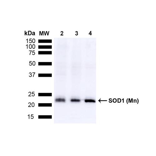
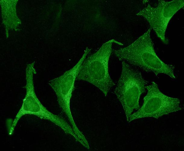
  • Immunocytochemistry/Immunofluorescence analysis using Rabbit Anti-SOD1 (Cu/Zn) Polyclonal Antibody (STPA00011). Tissue: Cervical cancer cell line (HeLa). Species: Human. Fixation: 2% Formaldehyde for 20 min at RT. Primary Antibody: Rabbit Anti-SOD1 (Cu/Zn) Polyclonal Antibody (STPA00011) at 1:120 for 12 hours at 4°C. Secondary Antibody: FITC Goat Anti-Rabbit (green) at 1:200 for 2 hours at RT. Western blot analysis of Human Cell line lysates showing detection of SOD1 (Cu/Zn) protein using Rabbit Anti-SOD1 (Cu/Zn) Polyclonal Antibody (STPA00011). Load: 15 µgprotein. Block: 1.5% BSA. Primary Antibody: Rabbit Anti-SOD1 (Cu/Zn) Polyclonal Antibody (STPA00011) at 1:1000 for 2 hours at RT. Secondary Antibody: Donkey Anti-Rabbit IgG: HRP for 1 hour at RT.

    Anti-SOD1 (Cu/Zn) Antibody

    SOD1 (Cu/Zn) AntibodySOD1 (Cu/Zn) Antibody is a rabbit polyclonal antibody directed against SOD1 (Cu/Zn), offered by Assay Genie for research applications. Reported applications include WB, IHC,...
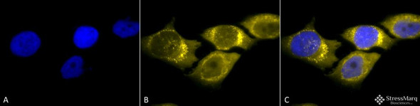
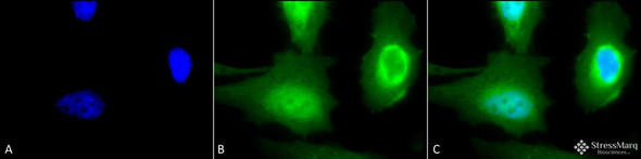
  • Immunocytochemistry/Immunofluorescence analysis using Rabbit Anti-PDI Polyclonal Antibody (STPA00010). Tissue: Cervical cancer cell line (HeLa). Species: Human. Fixation: 2% Formaldehyde for 20 min at RT. Primary Antibody: Rabbit Anti-PDI Polyclonal Antibody (STPA00010) at 1:100 for 12 hours at 4°C. Secondary Antibody: R-PE Goat Anti-Rabbit (yellow) at 1:200 for 2 hours at RT. Counterstain: DAPI (blue) nuclear stain at 1:40000 for 2 hours at RT. Western blot analysis of Rat tissue mix showing detection of PDI protein using Rabbit Anti-PDI Polyclonal Antibody (STPA00010). Load: 15 µgprotein. Block: 1.5% BSA. Primary Antibody: Rabbit Anti-PDI Polyclonal Antibody (STPA00010) at 1:4000 for 2 hours at RT. Secondary Antibody: Donkey Anti-Rabbit IgG: HRP for 1 hour at RT.

    Anti-PDI Antibody

    PDI AntibodyPDI Antibody is a rabbit polyclonal antibody directed against PDI, offered by Assay Genie for research applications. Reported applications include WB, IHC, ICC/IF and IP. Reported...
  • Immunocytochemistry/Immunofluorescence analysis using Rabbit Anti-HO-1 Polyclonal Antibody (STPA00009). Tissue: Heat Shocked Cervical cancer cell line (HeLa). Species: Human. Fixation: 2% Formaldehyde for 20 min at RT. Primary Antibody: Rabbit Anti-HO-1 Polyclonal Antibody (STPA00009) at 1:100 for 12 hours at 4°C. Secondary Antibody: FITC Goat Anti-Rabbit (green) at 1:200 for 2 hours at RT. Western blot analysis of Human Cell line lysates showing detection of HO-1 protein using Rabbit Anti-HO-1 Polyclonal Antibody (STPA00009). Load: 15 µgprotein. Block: 1.5% BSA. Primary Antibody: Rabbit Anti-HO-1 Polyclonal Antibody (STPA00009) at 1:1000 for 2 hours at RT. Secondary Antibody: Donkey Anti-Rabbit IgG: HRP for 1 hour at RT.

    Anti-HO-1 Antibody

    HO-1 AntibodyHO-1 Antibody is a rabbit polyclonal antibody directed against HO-1, offered by Assay Genie for research applications. Reported applications include WB, ICC/IF and IHC. Reported...
  • Immunocytochemistry/Immunofluorescence analysis using Rabbit Anti-KDEL Polyclonal Antibody (STPA00008). Tissue: Heat Shocked Cervical cancer cell line (HeLa). Species: Human. Fixation: 2% Formaldehyde for 20 min at RT. Primary Antibody: Rabbit Anti-KDEL Polyclonal Antibody (STPA00008) at 1:100 for 12 hours at 4°C. Secondary Antibody: FITC Goat Anti-Rabbit (green) at 1:200 for 2 hours at RT. Western blot analysis of Human Cell line lysates showing detection of KDEL protein using Rabbit Anti-KDEL Polyclonal Antibody (STPA00008). Primary Antibody: Rabbit Anti-KDEL Polyclonal Antibody (STPA00008) at 1:1000, 1:500.

    Anti-KDEL Antibody

    KDEL AntibodyKDEL Antibody is a rabbit polyclonal antibody directed against KDEL, offered by Assay Genie for research applications. Reported applications include WB, ICC/IF and IHC. Reported...
  • Immunocytochemistry/Immunofluorescence analysis using Rabbit Anti-Calnexin Polyclonal Antibody (STPA00007). Tissue: Cervical cancer cell line (HeLa). Species: Human. Primary Antibody: Rabbit Anti-Calnexin Polyclonal Antibody (STPA00007) at 1:100. Secondary Antibody: FITC Goat Anti-Rabbit (green). Western blot analysis of Human Cervical cancer cell line (HeLa) lysate showing detection of Calnexin protein using Rabbit Anti-Calnexin Polyclonal Antibody (STPA00007). Primary Antibody: Rabbit Anti-Calnexin Polyclonal Antibody (STPA00007) at 1:1000.

    Anti-Calnexin-CT Antibody

    Calnexin-CT AntibodyCalnexin-CT Antibody is a rabbit polyclonal antibody directed against Calnexin-CT, offered by Assay Genie for research applications. Reported applications include WB, ICC/IF and...