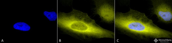
Anti-Rubicon Antibody

- SKU:
- STPA00067
- Antibody Type:
- Polyclonal
- Applications:
- WB
- Applications:
- ICC/IF
- Applications:
- IHC
- Reactivity:
- Human
- Host Species:
- Rabbit



Description
Rubicon Antibody
Rubicon Antibody is a rabbit polyclonal antibody directed against Rubicon, offered by Assay Genie for research applications. Reported applications include WB, ICC/IF and IHC. Reported reactivity: Human. Purified by Peptide Affinity.
Rubicon (RUN domain Beclin-1-interacting and cysteine-rich-containing protein) is a negative regulator of autophagy, a cellular degradation pathway essential for maintaining neuronal homeostasis. As neurons are particularly vulnerable to the accumulation of damaged proteins and organelles, dysregulation of autophagy is a hallmark of many neurodegenerative diseases, including Alzheimer’s, Parkinson’s, and ALS. Rubicon inhibits autophagosome maturation by interacting with components of the PI3K complex and RAB7A, thereby blocking lysosomal fusion. In the aging brain, elevated Rubicon levels have been associated with impaired autophagic flux and increased vulnerability to proteotoxic stress 1. Notably, Rubicon expression is upregulated in Alzheimer’s disease, where it contributes to amyloid precursor protein (APP) and amyloid-β (Aβ) accumulation. Neuronal overexpression of Rubicon suppresses Aβ clearance, while its knockdown enhances autophagic degradation and reduces extracellular Aβ deposition (1). Recent findings also highlight Rubicon’s role in mitophagy regulation. Phosphorylation of RAB7A at Ser72 disrupts its interaction with Rubicon, relieving autophagy inhibition and promoting mitochondrial quality control 2. This phospho-switch mechanism underscores Rubicon’s dynamic regulation in response to cellular stress. Given its central role in autophagy suppression and protein aggregation, Rubicon is a promising therapeutic target. Strategies aimed at modulating Rubicon expression or disrupting its inhibitory interactions may restore autophagic balance and mitigate neurodegenerative progression. This antibody is also available conjugated to ATTO 390, ATTO 488, ATTO 594, APC, Biotin, FITC, HRP, PerCP and RPE. Supplied in 100 µg. For research use only; not for diagnostic or therapeutic procedures.
| Product Name: | Rubicon Antibody |
| Product SKU: | STPA00067 |
| Size: | 100 µg |
| Target: | Rubicon |
| Host Species: | Rabbit |
| Antibody Type: | Polyclonal |
| Research Areas: | Cardiovascular System, Heart, Cancer, Autophagy, Cell Signaling, Metabolism, Metabolism processes |
| Buffer: | PBS, 50% glycerol, 0.09% sodium azide |
| Storage & Shipping: | Store at -20°C. Shipped on Blue Ice or 4°C. Conjugated variants should be stored according to the product label. |
| Format: | Liquid |
| Applications: | WB, ICC/IF, IHC |
| Purification: | Peptide Affinity Purified |
| Concentration: | 1 mg/ml |
| Reactivity: | Human |
| Recommended Dilution: | WB (1:1000), ICC/IF (1:100); IHC (1:50); optimal dilutions for assays should be determined by the user. |
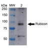
| Specificity: | Detects ~95 kDa. |
| Guarantee: | 12 months from date of dispatch |
| Immunogen: | Synthetic peptide from the N-terminal of Human Rubicon |
| Immunogen Species: | Human |
| Accession Number: | NP_001139114.1 |
| Gene ID: | 9711 |
| Swiss-Prot: | Q92622 |
| Cellular Localization: | Late Endosome | Lysosome | Early Endosome |
| Alternative Names: | Run domain Beclin-1-interacting and cysteine-rich domain-containing protein, KIAA0226, Rubicon, Baron, RUBCN, Beclin-1 associated RUN domain containing protein |
| Note: | For research use only. |
Related Products
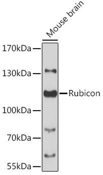
Rubicon Rabbit Polyclonal Antibody (CAB13169)
Autophagy Antibodies


Anti-HSP110 Antibody


Anti-NHE3 Antibody


Anti-Rab4 Antibody