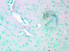
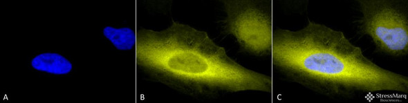
Cav1.2 Antibody [S57] is a mouse recombinant monoclonal antibody directed against Cav1.2, offered by Assay Genie for research applications. Reported applications include WB, IHC, ICC/IF, IP and AM. Reported reactivity: Human, Mouse, Rat and Hamster. Supplied as IgG1, purified by Protein G.
Cav1.2, encoded by the CACNA1C gene, is a major subunit of the L-type voltage-gated calcium channel family, best known for its role in cardiac excitation-contraction coupling. However, beyond the heart, Cav1.2 is highly expressed in the brain, where it regulates calcium influx critical for neuronal excitability, synaptic plasticity, and gene transcription. Dysregulation of Cav1.2 has been implicated in a range of neurological and neuropsychiatric disorders. Mutations in CACNA1C are causally linked to Timothy syndrome, a rare multisystem disorder characterized by cardiac arrhythmias, autism spectrum features, and cognitive impairment. Additionally, variants in CACNA1C have been associated with Brugada syndrome and are increasingly recognized as genetic risk factors for bipolar disorder, schizophrenia, and major depressive disorder. In the context of neurodegenerative disease, altered Cav1.2 function may contribute to disrupted calcium homeostasis, oxidative stress, and neuronal vulnerability—hallmarks of conditions such as Alzheimer’s and Parkinson’s disease. Its role in activity-dependent gene expression also positions Cav1.2 as a key modulator of long-term neuronal survival and plasticity. Given its dual importance in cardiac and neural physiology, Cav1.2 is a promising target for therapeutic strategies aimed at restoring calcium signaling balance in both neurodevelopmental and neurodegenerative disorders. This antibody is also available conjugated to ATTO 390, ATTO 488, ATTO 594, APC, Biotin, FITC, HRP, PerCP and RPE. Supplied in 100 µg. For research use only; not for diagnostic or therapeutic procedures.
HSP110 AntibodyHSP110 Antibody is a rabbit polyclonal antibody directed against HSP110, offered by Assay Genie for research applications. Reported applications include WB, IHC and ICC/IF. Reported...
NHE3 AntibodyNHE3 Antibody is a rabbit polyclonal antibody directed against NHE3, offered by Assay Genie for research applications. Reported applications include WB, ICC/IF and IHC. Reported...
Rab4 AntibodyRab4 Antibody is a rabbit polyclonal antibody directed against Rab4, offered by Assay Genie for research applications. Reported applications include WB, IHC and ICC/IF. Reported...
Ubiquitin AntibodyUbiquitin Antibody is a rabbit polyclonal antibody directed against Ubiquitin, offered by Assay Genie for research applications. Reported applications include WB, IHC, ICC/IF and IP...



![Western Blot analysis with Anti-Cav1.2 Antibody [S57] (STMA00080) showing detection of Cav1.2 in HEK293 whole cell lysate (Rockland W09-000-365) heated at 60°C for 10 minutes. Human Cav1.2 protein (UniProt Q13936) has a predicted molecular weight of ~249 kDa. Block: 2% skim milk + 2% BSA. Primary Antibody: Mouse Anti-Rabbit Cav1.2 Monoclonal IgG1 (STMA00080) at 1:1000 for 2 hours at RT.](https://cdn11.bigcommerce.com/s-rd6ounxcu2/images/stencil/1280x1280/products/120428/135238/anti-cav1.2-antibody-s57__35759__09626.1786857990.jpg?c=1&imbypass=on "Western Blot analysis with Anti-Cav1.2 Antibody [S57] (STMA00080) showing detection of Cav1.2 in HEK293 whole cell lysate (Rockland W09-000-365) heated at 60°C for 10 minutes. Human Cav1.2 protein (UniProt Q13936) has a predicted molecular weight of ~249 kDa. Block: 2% skim milk + 2% BSA. Primary Antibody: Mouse Anti-Rabbit Cav1.2 Monoclonal IgG1 (STMA00080) at 1:1000 for 2 hours at RT.")
![Western Blot analysis with Anti-Cav1.2 Antibody [S57] (STMA00080) showing detection of Cav1.2 in mouse brain lysate (Novus NB820-59657) heated at 60°C for 10 minutes. Mouse Cav1.2 protein (UniProt Q01815) has a predicted molecular weight of ~240 kDa. Block: 2% skim milk + 2% BSA. Primary Antibody: Mouse Anti-Rabbit Cav1.2 Monoclonal IgG1 (STMA00080) at 1:1000 for 2 hours at RT.](https://cdn11.bigcommerce.com/s-rd6ounxcu2/images/stencil/1280x1280/products/120428/135239/anti-cav1.2-antibody-s57__80268__22353.1786857992.jpg?c=1&imbypass=on "Western Blot analysis with Anti-Cav1.2 Antibody [S57] (STMA00080) showing detection of Cav1.2 in mouse brain lysate (Novus NB820-59657) heated at 60°C for 10 minutes. Mouse Cav1.2 protein (UniProt Q01815) has a predicted molecular weight of ~240 kDa. Block: 2% skim milk + 2% BSA. Primary Antibody: Mouse Anti-Rabbit Cav1.2 Monoclonal IgG1 (STMA00080) at 1:1000 for 2 hours at RT.")
![Western Blot analysis with Anti-Cav1.2 Antibody [S57] (STMA00080) showing detection of Cav1.2 in rat brain membrane lysate (prepared in-house) heated at 37°C for 10 minutes. Rat Cav1.2 protein (UniProt P22002) has a predicted molecular weight of ~243 kDa. Block: 2% skim milk + 2% BSA. Primary Antibody: Mouse Anti-Rabbit Cav1.2 Monoclonal IgG1 (STMA00080) at 1:1000 for 2 hours at RT.](https://cdn11.bigcommerce.com/s-rd6ounxcu2/images/stencil/1280x1280/products/120428/135241/anti-cav1.2-antibody-s57__25399__50413.1786857993.jpg?c=1&imbypass=on "Western Blot analysis with Anti-Cav1.2 Antibody [S57] (STMA00080) showing detection of Cav1.2 in rat brain membrane lysate (prepared in-house) heated at 37°C for 10 minutes. Rat Cav1.2 protein (UniProt P22002) has a predicted molecular weight of ~243 kDa. Block: 2% skim milk + 2% BSA. Primary Antibody: Mouse Anti-Rabbit Cav1.2 Monoclonal IgG1 (STMA00080) at 1:1000 for 2 hours at RT.")




![Western Blot analysis with Anti-Cav1.2 Antibody [S57] (STMA00080) showing detection of Cav1.2 in HEK293 whole cell lysate (Rockland W09-000-365) heated at 60°C for 10 minutes. Human Cav1.2 protein (UniProt Q13936) has a predicted molecular weight of ~249 kDa. Block: 2% skim milk + 2% BSA. Primary Antibody: Mouse Anti-Rabbit Cav1.2 Monoclonal IgG1 (STMA00080) at 1:1000 for 2 hours at RT.](https://cdn11.bigcommerce.com/s-rd6ounxcu2/images/stencil/100x100/products/120428/135238/anti-cav1.2-antibody-s57__35759__09626.1786857990.jpg?c=1 "Western Blot analysis with Anti-Cav1.2 Antibody [S57] (STMA00080) showing detection of Cav1.2 in HEK293 whole cell lysate (Rockland W09-000-365) heated at 60°C for 10 minutes. Human Cav1.2 protein (UniProt Q13936) has a predicted molecular weight of ~249 kDa. Block: 2% skim milk + 2% BSA. Primary Antibody: Mouse Anti-Rabbit Cav1.2 Monoclonal IgG1 (STMA00080) at 1:1000 for 2 hours at RT.")
![Western Blot analysis with Anti-Cav1.2 Antibody [S57] (STMA00080) showing detection of Cav1.2 in mouse brain lysate (Novus NB820-59657) heated at 60°C for 10 minutes. Mouse Cav1.2 protein (UniProt Q01815) has a predicted molecular weight of ~240 kDa. Block: 2% skim milk + 2% BSA. Primary Antibody: Mouse Anti-Rabbit Cav1.2 Monoclonal IgG1 (STMA00080) at 1:1000 for 2 hours at RT.](https://cdn11.bigcommerce.com/s-rd6ounxcu2/images/stencil/100x100/products/120428/135239/anti-cav1.2-antibody-s57__80268__22353.1786857992.jpg?c=1 "Western Blot analysis with Anti-Cav1.2 Antibody [S57] (STMA00080) showing detection of Cav1.2 in mouse brain lysate (Novus NB820-59657) heated at 60°C for 10 minutes. Mouse Cav1.2 protein (UniProt Q01815) has a predicted molecular weight of ~240 kDa. Block: 2% skim milk + 2% BSA. Primary Antibody: Mouse Anti-Rabbit Cav1.2 Monoclonal IgG1 (STMA00080) at 1:1000 for 2 hours at RT.")
![Western Blot analysis with Anti-Cav1.2 Antibody [S57] (STMA00080) showing detection of Cav1.2 in rat brain membrane lysate (prepared in-house) heated at 37°C for 10 minutes. Rat Cav1.2 protein (UniProt P22002) has a predicted molecular weight of ~243 kDa. Block: 2% skim milk + 2% BSA. Primary Antibody: Mouse Anti-Rabbit Cav1.2 Monoclonal IgG1 (STMA00080) at 1:1000 for 2 hours at RT.](https://cdn11.bigcommerce.com/s-rd6ounxcu2/images/stencil/100x100/products/120428/135241/anti-cav1.2-antibody-s57__25399__50413.1786857993.jpg?c=1 "Western Blot analysis with Anti-Cav1.2 Antibody [S57] (STMA00080) showing detection of Cav1.2 in rat brain membrane lysate (prepared in-house) heated at 37°C for 10 minutes. Rat Cav1.2 protein (UniProt P22002) has a predicted molecular weight of ~243 kDa. Block: 2% skim milk + 2% BSA. Primary Antibody: Mouse Anti-Rabbit Cav1.2 Monoclonal IgG1 (STMA00080) at 1:1000 for 2 hours at RT.")