Human TNFRSF1B (Tumor Necrosis Factor Receptor Superfamily, Member 1B) ELISA Kit
The Human TNFRSF1B (Tumor Necrosis Factor Receptor Superfamily Member 1B) ELISA Kit is a powerful tool for accurately measuring TNFRSF1B levels in human serum, plasma, and cell culture supernatants. This kit offers superior sensitivity and specificity, guaranteeing precise and consistent results that are essential for a variety of research applications.TNFRSF1B is a key receptor in the tumor necrosis factor pathway, playing a vital role in immune response, inflammation, and cell survival. Dysregulation of TNFRSF1B has been linked to various diseases, including autoimmune disorders, cancer, and inflammatory conditions, making it a valuable biomarker for studying these diseases and developing targeted therapies.With its reliable performance and comprehensive scope of applications, the Human TNFRSF1B ELISA Kit from Assay Genie is an indispensable tool for researchers seeking to unravel the complexities of TNFRSF1B signaling and its implications in human health and disease.
Product Name:
Human TNFRSF1B (Tumor Necrosis Factor Receptor Superfamily, Member 1B) ELISA Kit
SKU:
HUES03302
Size:
96 Assays
Detection Method:
Colorimetric method, ELISA, Sandwich
Assay type:
Sandwich-ELISA
Assay time:
3 h 30 min
Sensitivity:
4.69 pg/mL
Detection range:
7.81-500 pg/mL
Reovery:
80%-120%
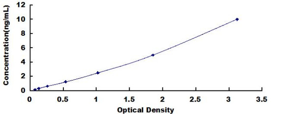
This ELISA kit uses the Sandwich-ELISA principle. The micro ELISA plate provided in this kit has been pre-coated with an antibody specific to the target protein. Standards or samples are added to the micro ELISA plate wells and bind to the immobilized antibody. A biotinylated detection antibody specific to the target protein is then added, followed by Avidin-Horseradish Peroxidase (HRP) conjugate. Free components are washed away. The substrate solution is added to each well, resulting in a color change. Only wells containing the target protein, detection antibody, and HRP conjugate will develop a blue color. The reaction is terminated by the addition of stop solution, resulting in a yellow color. The optical density (OD) is measured at 450 nm ± 2 nm. The OD value is directly proportional to the concentration of the target protein in the sample and is determined using a standard curve.