
Signal Transduction
-


Anti-Acetylated Lysine Antibody
Acetylated Lysine AntibodyAcetylated Lysine Antibody is a rabbit polyclonal antibody directed against Acetylated Lysine, offered by Assay Genie for research applications. Reported applications... -
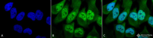

Anti-Phosphothreonine Antibody
Phosphothreonine AntibodyPhosphothreonine Antibody is a rabbit polyclonal antibody directed against Phosphothreonine, offered by Assay Genie for research applications. Reported applications include... -

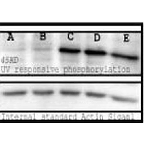
Anti-Phosphoserine Antibody
Phosphoserine AntibodyPhosphoserine Antibody is a rabbit polyclonal antibody directed against Phosphoserine, offered by Assay Genie for research applications. Reported applications include WB, IHC,... -
![Western blot analysis with Rabbit Anti-Rat KChIP3 Monoclonal Antibody, Clone X12 (STMA00233) showing detection of Human KChIP3 protein in a transient over-expression lysate (OriGene LY415545). Block: 2% skim milk + 2% BSA for 1 hour at RT. Primary Antibody: Rabbit Anti-Rat KChIP3 Recombinant Monoclonal [X12] (STMA00233) at 1:1000 for 2 hours at RT. Secondary Antibody: Goat anti-rabbit IgG-HRP (H&L) at 1:5000 for 1 hour at RT.](https://cdn11.bigcommerce.com/s-rd6ounxcu2/images/stencil/590x590/products/120586/135674/anti-kchip3-antibody-x12__45632__79049.1786858497.jpg?c=1 "Western blot analysis with Rabbit Anti-Rat KChIP3 Monoclonal Antibody, Clone X12 (STMA00233) showing detection of Human KChIP3 protein in a transient over-expression lysate (OriGene LY415545). Block: 2% skim milk + 2% BSA for 1 hour at RT. Primary Antibody: Rabbit Anti-Rat KChIP3 Recombinant Monoclonal [X12] (STMA00233) at 1:1000 for 2 hours at RT. Secondary Antibody: Goat anti-rabbit IgG-HRP (H&L) at 1:5000 for 1 hour at RT.")
![Western blot analysis with Rabbit Anti-Rat KChIP3 Monoclonal Antibody, Clone X12 (STMA00233) showing detection of Rat KChIP3 protein in a Rat brain membrane lysate. Block: 2% skim milk + 2% BSA for 1 hour at RT. Primary Antibody: Rabbit Anti-Rat KChIP3 Recombinant Monoclonal [X12] (STMA00233) at 1:1000 for 2 hours at RT. Secondary Antibody: Goat anti-rabbit IgG-HRP (H&L) at 1:5000 for 1 hour at RT.](https://cdn11.bigcommerce.com/s-rd6ounxcu2/images/stencil/590x590/products/120586/135676/anti-kchip3-antibody-x12__09538__24764.1786858499.jpg?c=1 "Western blot analysis with Rabbit Anti-Rat KChIP3 Monoclonal Antibody, Clone X12 (STMA00233) showing detection of Rat KChIP3 protein in a Rat brain membrane lysate. Block: 2% skim milk + 2% BSA for 1 hour at RT. Primary Antibody: Rabbit Anti-Rat KChIP3 Recombinant Monoclonal [X12] (STMA00233) at 1:1000 for 2 hours at RT. Secondary Antibody: Goat anti-rabbit IgG-HRP (H&L) at 1:5000 for 1 hour at RT.")
Anti-KChIP3 Antibody [X12]
KChIP3 Antibody [X12]KChIP3 Antibody [X12] is a rabbit recombinant monoclonal antibody directed against KChIP3, offered by Assay Genie for research applications. Reported applications include WB... -
![Western blot analysis with Rabbit Anti-Rat SAP97 Monoclonal Antibody, Clone G28 (STMA00235) showing detection of Rat SAP97 protein in a Rat brain membrane lysate. Block: 2% skim milk + 2% BSA for 1 hour at RT. Primary Antibody: Rabbit Anti-Rat SAP97 Recombinant Monoclonal [G28] (STMA00235) at 1:1000 for 2 hours at RT. Secondary Antibody: Goat anti-rabbit IgG-HRP (H&L) at 1:5000 for 1 hour at RT.](https://cdn11.bigcommerce.com/s-rd6ounxcu2/images/stencil/590x590/products/120585/135671/anti-sap97-antibody-g28__65287__23952.1786858496.jpg?c=1 "Western blot analysis with Rabbit Anti-Rat SAP97 Monoclonal Antibody, Clone G28 (STMA00235) showing detection of Rat SAP97 protein in a Rat brain membrane lysate. Block: 2% skim milk + 2% BSA for 1 hour at RT. Primary Antibody: Rabbit Anti-Rat SAP97 Recombinant Monoclonal [G28] (STMA00235) at 1:1000 for 2 hours at RT. Secondary Antibody: Goat anti-rabbit IgG-HRP (H&L) at 1:5000 for 1 hour at RT.")
![Immunocytochemistry/Immunofluorescence analysis with Rabbit Anti-Rat SAP97 Monoclonal Antibody, Clone G28 (STMA00235). Tissue: Pheochromocytoma cells (PC-12). Species: Rat. Fixation: 4% PFA for 10 min at RT. Permeabilization: 0.15% TritonX for 15 min at RT. Blocking: 10% goat serum for 40 min at RT. Primary Antibody: Rabbit Anti-Rat SAP97 Recombinant Monoclonal [G28] (STMA00235) at 1:100 for 1 hour at RT.](https://cdn11.bigcommerce.com/s-rd6ounxcu2/images/stencil/590x590/products/120585/135675/anti-sap97-antibody-g28__08062__38284.1786858498.jpg?c=1 "Immunocytochemistry/Immunofluorescence analysis with Rabbit Anti-Rat SAP97 Monoclonal Antibody, Clone G28 (STMA00235). Tissue: Pheochromocytoma cells (PC-12). Species: Rat. Fixation: 4% PFA for 10 min at RT. Permeabilization: 0.15% TritonX for 15 min at RT. Blocking: 10% goat serum for 40 min at RT. Primary Antibody: Rabbit Anti-Rat SAP97 Recombinant Monoclonal [G28] (STMA00235) at 1:100 for 1 hour at RT.")
Anti-SAP97 Antibody [G28]
SAP97 Antibody [G28]SAP97 Antibody [G28] is a rabbit recombinant monoclonal antibody directed against SAP97, offered by Assay Genie for research applications. Reported applications include WB and... -
![Western blot analysis with Rabbit Anti- Slo1 Monoclonal Antibody, Clone S22 (STMA00229) showing detection of Human Slo1 protein in a transient over-expression (OE) lysate (OriGene LY400815). Block: 2% skim milk + 2% BSA for 1 hour at RT. Primary Antibody: Rabbit Anti-Mouse Slo1 Recombinant Monoclonal [S22] (STMA00229) at 1:1000 for 2 hours at RT. Secondary Antibody: at 1:5000 for 1 hour at RT.](https://cdn11.bigcommerce.com/s-rd6ounxcu2/images/stencil/590x590/products/120580/135685/anti-slo1-antibody-s22__47681__58357.1786858507.jpg?c=1 "Western blot analysis with Rabbit Anti- Slo1 Monoclonal Antibody, Clone S22 (STMA00229) showing detection of Human Slo1 protein in a transient over-expression (OE) lysate (OriGene LY400815). Block: 2% skim milk + 2% BSA for 1 hour at RT. Primary Antibody: Rabbit Anti-Mouse Slo1 Recombinant Monoclonal [S22] (STMA00229) at 1:1000 for 2 hours at RT. Secondary Antibody: at 1:5000 for 1 hour at RT.")
![Immunocytochemistry/Immunofluorescence analysis with Rabbit Anti- Slo1 Monoclonal Antibody, Clone S22 (STMA00229). Tissue: Neuroblastoma cells (Kelly). Species: Human. Fixation: 4% PFA for 10 min at RT. Permeabilization: 0.15% TritonX for 15 min at RT. Blocking: 10% goat serum for 40 min at RT. Primary Antibody: Rabbit Anti-Mouse Slo1 Recombinant Monoclonal [S22] (STMA00229) at 1:100 for 1 hour at RT.](https://cdn11.bigcommerce.com/s-rd6ounxcu2/images/stencil/590x590/products/120580/135688/anti-slo1-antibody-s22__70032__85884.1786858513.jpg?c=1 "Immunocytochemistry/Immunofluorescence analysis with Rabbit Anti- Slo1 Monoclonal Antibody, Clone S22 (STMA00229). Tissue: Neuroblastoma cells (Kelly). Species: Human. Fixation: 4% PFA for 10 min at RT. Permeabilization: 0.15% TritonX for 15 min at RT. Blocking: 10% goat serum for 40 min at RT. Primary Antibody: Rabbit Anti-Mouse Slo1 Recombinant Monoclonal [S22] (STMA00229) at 1:100 for 1 hour at RT.")
Anti-Slo1 Antibody [S22]
Slo1 Antibody [S22]Slo1 Antibody [S22] is a rabbit recombinant monoclonal antibody directed against Slo1, offered by Assay Genie for research applications. Reported applications include WB, IHC and... -
![Western blot analysis with Rabbit Anti-Kv1.2 Monoclonal Antibody, Clone C25 (STMA00232) showing detection of Human Kv1.2 protein in a transient over-expression (OE) lysate (OriGene LY401550). Block: 2% skim milk + 2% BSA for 1 hour at RT. Primary Antibody: Rabbit Anti-Human Kv1.2 Recombinant Monoclonal [C25] (STMA00232) at 1:10000 for 2 hours at RT. Secondary Antibody: Goat anti-Rabbit IgG-HRP (H&L) at 1:5000 for 1 hour at RT.](https://cdn11.bigcommerce.com/s-rd6ounxcu2/images/stencil/590x590/products/120579/135689/anti-kv1.2-antibody-c25__57216__69561.1786858513.jpg?c=1 "Western blot analysis with Rabbit Anti-Kv1.2 Monoclonal Antibody, Clone C25 (STMA00232) showing detection of Human Kv1.2 protein in a transient over-expression (OE) lysate (OriGene LY401550). Block: 2% skim milk + 2% BSA for 1 hour at RT. Primary Antibody: Rabbit Anti-Human Kv1.2 Recombinant Monoclonal [C25] (STMA00232) at 1:10000 for 2 hours at RT. Secondary Antibody: Goat anti-Rabbit IgG-HRP (H&L) at 1:5000 for 1 hour at RT.")
![Immunocytochemistry/Immunofluorescence analysis with Rabbit Anti-Kv1.2 Monoclonal Antibody, Clone C25 (STMA00232). Tissue: Pheochromocytoma cells (PC-12). Species: Rat. Fixation: 4% PFA for 10 min at RT. Permeabilization: 0.15% TritonX for 15 min at RT. Blocking: 10% goat serum for 40 min at RT. Primary Antibody: Rabbit Anti-Human Kv1.2 Recombinant Monoclonal [C25] (STMA00232) at 1:100 for 1 hour at RT.](https://cdn11.bigcommerce.com/s-rd6ounxcu2/images/stencil/590x590/products/120579/135694/anti-kv1.2-antibody-c25__20109__19634.1786858514.jpg?c=1 "Immunocytochemistry/Immunofluorescence analysis with Rabbit Anti-Kv1.2 Monoclonal Antibody, Clone C25 (STMA00232). Tissue: Pheochromocytoma cells (PC-12). Species: Rat. Fixation: 4% PFA for 10 min at RT. Permeabilization: 0.15% TritonX for 15 min at RT. Blocking: 10% goat serum for 40 min at RT. Primary Antibody: Rabbit Anti-Human Kv1.2 Recombinant Monoclonal [C25] (STMA00232) at 1:100 for 1 hour at RT.")
Anti-Kv1.2 Antibody [C25]
Kv1.2 Antibody [C25]Kv1.2 Antibody [C25] is a rabbit recombinant monoclonal antibody directed against Kv1.2, offered by Assay Genie for research applications. Reported applications include WB, IHC... -
![Western blot analysis with Rabbit Anti-Kv1.3 Monoclonal Antibody, Clone CAR1.1 (STMA00234) showing detection of Human Kv1.3 protein in a transient over-expression (OE) lysate (OriGene LY4194581). Block: 2% skim milk + 2% BSA for 1 hour at RT. Primary Antibody: Rabbit Anti-Rat Kv1.3 Recombinant Monoclonal [CAR1.1] (STMA00234) at 1:10,000 for 2 hours at RT. Secondary Antibody: Goat anti-rabbit IgG-HRP (H&L) at 1:5000 for 1 hour at RT.](https://cdn11.bigcommerce.com/s-rd6ounxcu2/images/stencil/590x590/products/120577/135686/anti-kv1.3-antibody-car1.1__63339__02077.1786858511.jpg?c=1 "Western blot analysis with Rabbit Anti-Kv1.3 Monoclonal Antibody, Clone CAR1.1 (STMA00234) showing detection of Human Kv1.3 protein in a transient over-expression (OE) lysate (OriGene LY4194581). Block: 2% skim milk + 2% BSA for 1 hour at RT. Primary Antibody: Rabbit Anti-Rat Kv1.3 Recombinant Monoclonal [CAR1.1] (STMA00234) at 1:10,000 for 2 hours at RT. Secondary Antibody: Goat anti-rabbit IgG-HRP (H&L) at 1:5000 for 1 hour at RT.")
![Immunocytochemistry/Immunofluorescence analysis with Rabbit Anti-Kv1.3 Monoclonal Antibody, Clone CAR1.1 (STMA00234). Tissue: Pheochromocytoma cells (PC-12). Species: Rat. Fixation: 4% PFA for 10 min at RT. Permeabilization: 0.15% TritonX for 15 min at RT. Blocking: 10% goat serum for 40 min at RT. Primary Antibody: Rabbit Anti-Rat Kv1.3 Recombinant Monoclonal [CAR1.1] (STMA00234) at 1:100 for 1 hour at RT.](https://cdn11.bigcommerce.com/s-rd6ounxcu2/images/stencil/590x590/products/120577/135687/anti-kv1.3-antibody-car1.1__76004__48333.1786858512.jpg?c=1 "Immunocytochemistry/Immunofluorescence analysis with Rabbit Anti-Kv1.3 Monoclonal Antibody, Clone CAR1.1 (STMA00234). Tissue: Pheochromocytoma cells (PC-12). Species: Rat. Fixation: 4% PFA for 10 min at RT. Permeabilization: 0.15% TritonX for 15 min at RT. Blocking: 10% goat serum for 40 min at RT. Primary Antibody: Rabbit Anti-Rat Kv1.3 Recombinant Monoclonal [CAR1.1] (STMA00234) at 1:100 for 1 hour at RT.")
Anti-Kv1.3 Antibody [CAR1.1]
Kv1.3 Antibody [CAR1.1]Kv1.3 Antibody [CAR1.1] is a rabbit recombinant monoclonal antibody directed against Kv1.3, offered by Assay Genie for research applications. Reported applications include WB,... -

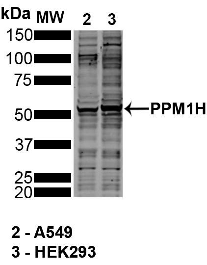
Anti-PPM1H Antibody [10D2]
PPM1H Antibody [10D2]PPM1H Antibody [10D2] is a mouse monoclonal antibody directed against PPM1H, offered by Assay Genie for research applications. Reported applications include WB and ICC/IF... -

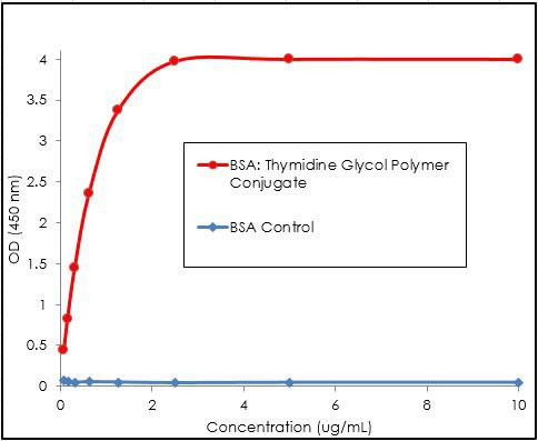
Anti-Thymidine glycol Antibody [B29]
Thymidine glycol Antibody [B29]Thymidine glycol Antibody [B29] is a rabbit monoclonal antibody directed against Thymidine glycol, offered by Assay Genie for research applications. Reported... -


Anti-Sulfotyrosine Antibody [12B11 (Formerly sold as 7C5)]
Sulfotyrosine Antibody [12B11 (Formerly sold as 7C5)]Sulfotyrosine Antibody [12B11 (Formerly sold as 7C5)] is a mouse recombinant monoclonal antibody directed against Sulfotyrosine, offered by Assay... -


Anti-Dityrosine Antibody [10A6]
Dityrosine Antibody [10A6]Dityrosine Antibody [10A6] is a mouse monoclonal antibody directed against Dityrosine, offered by Assay Genie for research applications. Reported applications include WB,...