Description
| Antibody Name: | Anti-RAB8A Antibody (CAB20976) |
| Antibody SKU: | CAB20976 |
| Antibody Size: | 50µL, 100µL |
| Application: | Western blotting, Immunofluorescence |
| Reactivity: | Human, Mouse, Rat |
| Host Species: | Rabbit |
| Immunogen: | Recombinant protein of Human RAB8A. |
| Application: | Western blotting, Immunofluorescence |
| Recommended Dilution: | WB 1:500 - 1:2000 IF 1:50 - 1:200 |
| Reactivity: | Human, Mouse, Rat |
| Positive Samples: | HeLa, HCT116, Mouse brain, Mouse thymus, Rat spinal cord |
| Immunogen: | Recombinant protein of Human RAB8A. |
| Purification Method: | Affinity purification |
| Storage Buffer: | Store at -20°C. Avoid freeze / thaw cycles. Buffer: PBS with 0.02% sodium azide, 0.05% BSA, 50% glycerol, pH7.3. |
| Isotype: | IgG |
| Sequence: | Email for sequence |
| Cellular Location: | Cell membrane, Cell projection, Cytoplasm, Cytoplasmic side, Cytoplasmic vesicle, Golgi apparatus, Lipid-anchor, Recycling endosome membrane, centriole, centrosome, cilium, cilium basal body, cytoskeleton, microtubule organizing center, phagosome, phagosome membrane |
| Calculated MW: | 23kDa |
| Observed MW: | 24KDa |
| Synonyms: | RAB8A, MEL, RAB8 |
| Background: | The protein encoded by this gene is a member of the RAS superfamily which are small GTP/GDP-binding proteins with an average size of 200 amino acids. The RAS-related proteins of the RAB/YPT family may play a role in the transport of proteins from the endoplasmic reticulum to the Golgi and the plasma membrane. This protein shares 97%, 96%, and 51% similarity with the dog RAB8, mouse MEL, and mouse YPT1 proteins, respectively and contains the 4 GTP/GDP-binding sites that are present in all the RAS proteins. The putative effector-binding site of this protein is similar to that of the RAB/YPT proteins. However, this protein contains a C-terminal CAAX motif that is characteristic of many RAS superfamily members but which is not found in YPT1 and the majority of RAB proteins. Although this gene was isolated as a transforming gene from a melanoma cell line, no linkage between MEL and malignant melanoma has been demonstrable. This oncogene is located 800 kb distal to MY09B on chromosome 19p13.1. |
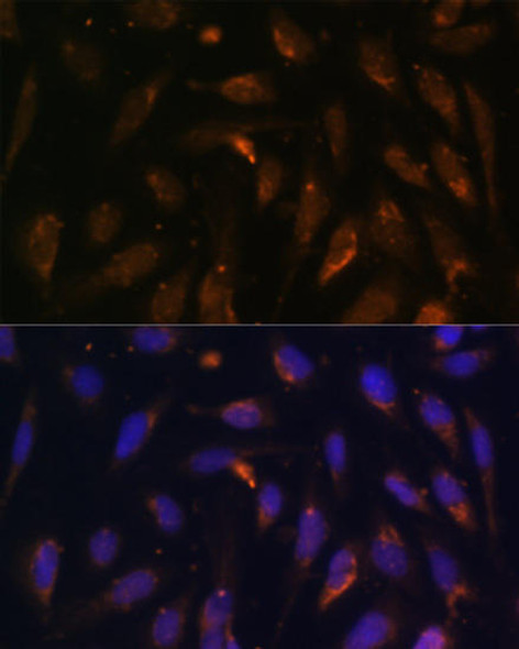
 | Immunofluorescence analysis of PC-12 cells using RAB8A Rabbit mAb at dilution of 1:100 (40x lens). Blue: DAPI for nuclear staining. |
 | Western blot analysis of extracts of Rat spinal cord, using RAB8A antibody at 1:1000 dilution. Secondary antibody: HRP Goat Anti-Rabbit IgG (H+L) at 1:10000 dilution. Lysates/proteins: 25ug per lane. Blocking buffer: 3% nonfat dry milk in TBST. Detection: ECL Basic Kit. Exposure time: 60s. |
 | Immunofluorescence analysis of NIH/3T3 cells using RAB8A Rabbit mAb at dilution of 1:100 (40x lens). Blue: DAPI for nuclear staining. |
 | Western blot analysis of extracts of various cell lines, using RAB8A antibody at 1:1000 dilution. Secondary antibody: HRP Goat Anti-Rabbit IgG (H+L) at 1:10000 dilution. Lysates/proteins: 25ug per lane. Blocking buffer: 3% nonfat dry milk in TBST. Detection: ECL Basic Kit. Exposure time: 3s. |


