Cav3.2 Antibody [N55/10 (Formerly sold as S55-10)]
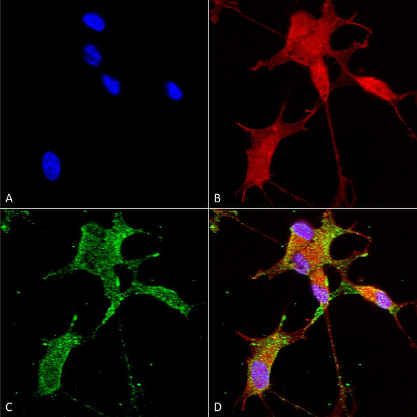
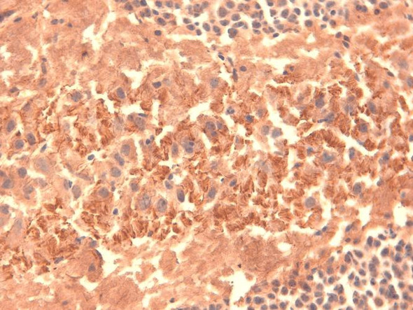
Cav3.2 Antibody [N55/10 (Formerly sold as S55-10)] is a mouse monoclonal antibody directed against Cav3.2, offered by Assay Genie for research applications. Reported applications include WB, IHC, ICC/IF, IP and AM. Reported reactivity: Human, Mouse and Rat. Supplied as IgG1, purified by Protein G.
Cav3.2, encoded by the CACNA1H gene, is a low-voltage-activated T-type calcium channel that plays a critical role in regulating neuronal excitability, rhythmic firing, and calcium homeostasis. It is widely expressed in the brain and peripheral tissues, where it contributes to burst firing in thalamic neurons and modulates sensory signal transmission. Genetic studies have linked mutations in CACNA1H to childhood absence epilepsy, highlighting Cav3.2’s role in thalamocortical oscillations and seizure susceptibility. Beyond epilepsy, dysregulation of Cav3.2 expression and function has been associated with broader neurological and neurodegenerative conditions, including neuropathic pain, Parkinson’s disease, and Alzheimer’s disease. Its involvement in calcium dysregulation—a hallmark of neurodegeneration—positions Cav3.2 as a potential therapeutic target for modulating neuronal survival and excitability. Interestingly, Cav3.2 is also implicated in non-neuronal pathologies, such as prostate cancer progression, where its upregulation may support androgen-independent tumor growth. This underscores the channel’s broader physiological relevance and potential as a drug target across multiple disease domains. Despite its clinical significance, many aspects of Cav3.2’s in vivo function, regulatory mechanisms, and disease-specific roles remain under investigation. Advancing our understanding of Cav3.2 could open new avenues for targeted therapies in epilepsy, neurodegeneration, and beyond. This antibody is also available conjugated to ATTO 390, ATTO 488, ATTO 594, APC, Biotin, FITC, HRP, PerCP and RPE. Supplied in 100 µg. For research use only; not for diagnostic or therapeutic procedures.
Product Name:
Cav3.2 Antibody [N55/10 (Formerly sold as S55-10)]
HCN4 Antibody [N114/10 (Formerly sold as S114-10)]HCN4 Antibody [N114/10 (Formerly sold as S114-10)] is a mouse recombinant monoclonal antibody directed against HCN4, offered by Assay Genie for...
KCNQ1 Antibody [N37A/10 (Formerly sold as S37A-10)]KCNQ1 Antibody [N37A/10 (Formerly sold as S37A-10)] is a mouse monoclonal antibody directed against KCNQ1, offered by Assay Genie for research...
Ankyrin R Antibody [N388A/10 (Formerly sold as S388A-10)]Ankyrin R Antibody [N388A/10 (Formerly sold as S388A-10)] is a mouse monoclonal antibody directed against Ankyrin R, offered by Assay Genie...
Sulfotyrosine Antibody [12B11 (Formerly sold as 7C5)]Sulfotyrosine Antibody [12B11 (Formerly sold as 7C5)] is a mouse recombinant monoclonal antibody directed against Sulfotyrosine, offered by Assay...
SUR1 Antibody [N289/16 (Formerly sold as S289-16)]SUR1 Antibody [N289/16 (Formerly sold as S289-16)] is a mouse monoclonal antibody directed against SUR1, offered by Assay Genie for research...