Therapeutic Drug Monitoring
Trastuzumab ELISA Kit (Herceptin®/Herclon®) - Free drug
- SKU:
- HUMB00030
- Product Type:
- ELISA Kit
- ELISA Type:
- Biosimilar ELISA
- Applications:
- ELISA
- Reactivity:
- Human
Description
Trastuzumab (Herceptin®, Herclon®) (Free drug) ELISA Kit
Enzyme immunoassay for the quantitative determination of free Trastuzumab (Herclon®, Herceptin®) in serum and plasma. The Assay Genie trastuzumab ELISA has been developed for the quantitative analysis of free trastuzumab in serum and plasma samples at high specificity.
Trastuzumab (Herceptin®, Herclon®) (Free drug) ELISA Kit Test Principle
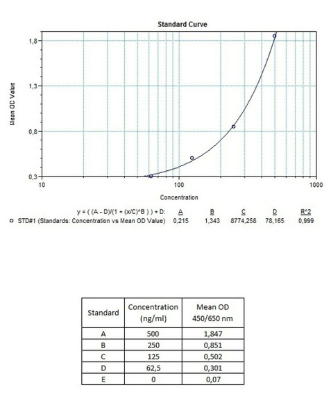
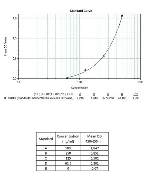
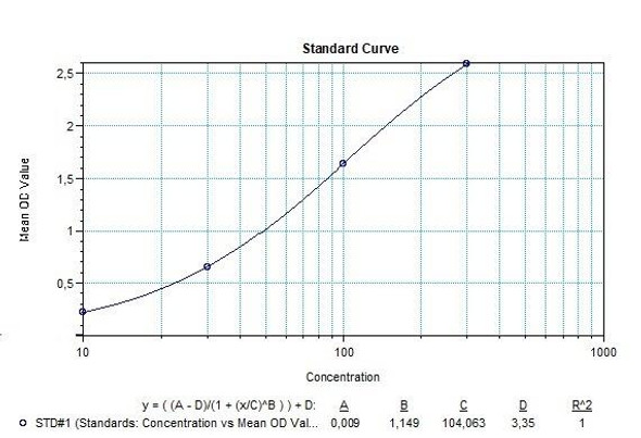
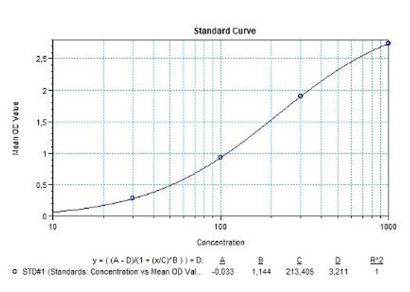
Solid phase enzyme-linked immunosorbent assay (ELISA) based on the sandwich principle. Standards and samples (serum or plasma) are incubated in the microtiter plate coated with the reactant for trastuzumab. After incubation, the wells are washed. Then, horse radish peroxidase (HRP) conjugated probe is added and binds to trastuzumab captured by the reactant on the surface of the wells. Following incubation wells are washed and the bound enzymatic activity is detected by addition of tetramethylbenzidine (TMB) chromogen substrate. Finally, the reaction is terminated with an acidic stop solution. The colour developed is proportional to the amount of trastuzumab in the sample or standard. Results of samples can be determined directly using the standard curve.
Trastuzumab (Herceptin®, Herclon®) (Free drug) ELISA Product Information
| Information | Description |
| Application | Free drug |
| Required Volume (µl) | 10 |
| Total Time (min) | 70 |
| Sample Typle | Serum, Plasma |
| Number of Assays | 96 |
| Detection Limit (ng/mL) | 2 |
| Recovery | 85-115% |
| Shelf Life (year) | 1 |
Trastuzumab (Herceptin®, Herclon®) (Free drug) ELISA - Key Information
Trastuzumab (Herceptin®, Herclon®) Mode of Action
Trastuzumab is a recombinant humanized IgG1 monoclonal antibody against the HER-2 receptor, a member of the epidermal growth factor receptors which is a photo-oncogene. Over-expressed in breast tumour cells, HER-2 overamplifies the signal provided by other receptors of the HER family by forming heterodimers.
The HER-2 receptor is a transmembrane tyrosine kinase receptor that consists of an extracellular ligand-binding domain, a transmembrane region, and an intracellular or cytoplasmic tyrosine kinase domain. It is activated by the formation of homodimers or heterodimers with other EGFR proteins, leading to dimerization and autophosphorylation and/or transphosphorylation of specific tyrosine residues in EGFR intracellular domains. Further downstream molecular signalling cascades are activated, such as the Ras/Raf/mitogen-activated protein kinase (MAPK), the phosphoinositide 3-kinase/Akt, and the phospholipase Cγ (PLCγ)/protein kinase C (PKC) pathways that promote cell growth and survival and cell cycle progression. Due to upregulation of HER-2 in tumour cells, hyperactivation of these signalling pathways and abnormal cell proliferation is observed. Trastuzumab binds to the extracellular ligandbinding domain and blocks the cleavage of the extracellular domain of HER-2 to induce its antibody-induced receptor downmodulation, and subsequently inhibits HER-2-mediated intracellular signalling cascades. Inhibition of MAPK and PI3K/Akt pathways lead to an increase in cell cycle arrest, and the suppression of cell growth and proliferation.
Trastuzumab also mediates the activation of antibody-dependent cell-mediated cytotoxicity (ADCC) by attracting the immune cells, such as natural killer (NK) cells, to tumour sites that overexpress HER-2. Intrinsic trastuzumab resistance has been noted for some patients with HER-2 positive breast cancer. Mechanisms involving trastuzumab resistance include deficiency of phosphatase and tensin homologue and activation of phosphoinositide 3- kinase, and the overexpression of other surface receptors, such as insulin-like growth factor.
Trastuzumab (Herceptin®, Herclon®) (Free drug) ELISA Kit Contents
| Size | Kit Contents |
| 1x12x8 | Microtiter Plate Break apart strips. Microtiter plate with 12 rows each of 8 wells coated with reactant. |
| 0,3 mL (each) | Standards A-E (10x) Standard A: 3000 ng/mL Standard B: 1000 ng/mL Standard C: 300 ng/mL Standard D: 100 ng/mL Standard E: 0 ng/mL Used for the standard curve and control. Contains trastuzumab, human serum and stabilizer, <0,1% NaN3. Standards are prepared concentrated (10x). |
| 0,3 mL (each | Control low and high levels (10x) Contains human serum and stabilizer, <0,1% NaN3. Controls are prepared concentrated (10x). |
| 2x50 mL | Assay buffer Ready to use. Blue coloured. Contains proteins, <0,1% NaN3. |
| 1x12 mL | Horse radish peroxidase conjugated probe. Ready to use. Red coloured. Contains HRP conjugated probe, stabilizer and preservatives. |
| 1x12 mL | TMB substrate solution. Ready to use. Contains 3,3′,5,5′- Tetramethylbenzidine (TMB). |
| 1x12mL | TMB stop solution. Ready to use. 1N HCl |
| 1x50mL | Wash buffer (20x). Prepared concentrated (20x) and should be diluted with the dilution rate given in the “Pretest setup instructions” before the test. Contains buffer with tween 20. |
| 2x1 | Adhesive Foil. For covering microtiter plate during incubation |
Trastuzumab (Herceptin®, Herclon®) (Free drug) ELISA Protocol
| 1 | Pipette 100µl of Assay Buffer non-exceptionally into each of the wells to be used. |
| 2 | Pipette 100 µL of each diluted Standards, Low level control, High level control and samples into the respective wells of microtiter plate Wells A1: Standard A B1: Standard B C1: Standard C D1: Standard D E1: Standard E F1: Low level control G1: High level control H1 and on: Samples |
| 3 | Cover the plate with adhesive foil. Briefly mix contents by gently shaking the plate. Incubate 30 minutes at room temperature (18-25°C) |
| 4 | Remove adhesive foil. Discard incubation solution. Wash plate three times each with 300 µL Wash Buffer. Remove excess solution by tapping the inverted plate on a paper towel |
| 5 | Pipette 100 µL Conjugate into each well |
| 6 | Cover the plate with adhesive foil. Incubate 30 minutes at room temperature (18-25°C) |
| 7 | Remove adhesive foil. Discard incubation solution Wash plate three times each with 300 µL Wash Buffer. Remove excess solution by tapping the inverted plate on a paper towel |
| 8 | Pipette 100 µL Substrate into each well |
| 9 | Incubate 10 minutes without adhesive foil at room temperature (18-25°C) in the dark |
| 10 | Stop the substrate reaction by adding 100 µL Stop Solution into each well. Briefly mix contents by gently shaking the plate. Colour changes from blue to yellow. |
| 11 | Measure optical density with a photometer at OD 450nm with reference wavelength 650 nm (450/650 nm) within 30 minutes after pipetting the Stop Solution. |


