Description
| Product Name: | QuickStep Mouse IgG (Immunoglobulin G) ELISA Kit |
| Product Code: | AEES00649 |
| Assay Type: | Sandwich |
| Format: | 96 Assays |
| Assay Time: | 2.5h |
| Reactivity: | Mouse |
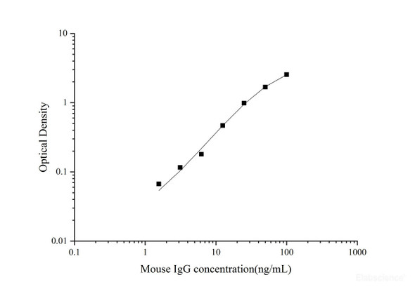
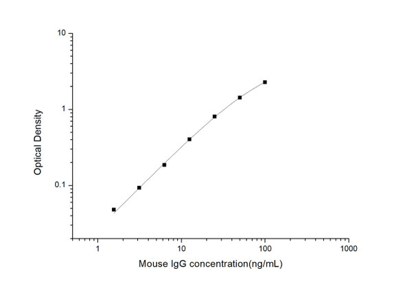
| Detection Range: | 1.56-100 ng/mL |
| Sensitivity: | 0.17 ng/mL |
| Sample Type & Sample Volume: | Serum and plasma, 50μL |
| Specificity: | This kit recognizes Mouse IgG in samples. No significant or interference between Mouse IgG and analogues was observed. |
| Reproducibility: | Both intra-CV and inter-CV are < 10%. |
| Application: | This ELISA kit applies to the in vitro quantitative determination of Mouse IgG concentrations in serum and plasma.Please consult technical support for the applicability if other biological fluids need to be tested. |
This ELISA kit uses the Sandwich-ELISA principle. The micro ELISA plate provided in this kit has been pre-coated with an antibody specific to Mouse IgG. Samples (or Standards) and biotinylated detection antibody specific for Mouse IgG are added to the micro ELISA plate wells. Mouse IgG would combine with the specific antibody. Then Avidin-Horseradish Peroxidase (HRP) conjugate are added successively to each micro plate well and incubated. Free components are washed away. The substrate solution is added to each well. Only those wells that contain Mouse IgG, biotinylated detection antibody and Avidin-HRP conjugate will appear blue in color. The enzyme-substrate reaction is terminated by the addition of stop solution and the color turns yellow. The optical density (OD) is measured spectrophotometrically at a wavelength of 450 ± 2 nm. The OD value is proportional to the concentration of Mouse IgG. You can calculate the concentration of Mouse IgG in the samples by comparing the OD of the samples to the standard curve.
| Kit Components: | An unopened kit can be stored at 2-8℃ for six months. After test, the unused wells and reagents should be stored according to the table below.
|
| 1. | Add 50μL standard or sample to the wells, immediately add 50μL Biotinylated Detection Ab working solution to each well. Incubate for 90 min at 37°C |
| 2. | Aspirate and wash the plate for 3 times |
| 3. | Add 100μL HRP conjugate working solution. Incubate for 30 min at 37°C. Aspirate and wash the plate for 5 times |
| 4. | Add 90μL Substrate Reagent. Incubate for 15 min at 37°C |
| 5. | Add 50μL Stop Solution |
| 6. | Read the plate at 450nm immediately. Calculation of the results |
Immunoglobulin G (IgG) is a class of immunoglobulins found in all body fluids. They are the smallest but most common antibodies (75 percent to 80 percent) in the body. IgG antibodies are large molecules of about 150 kDa[1], made of four peptide chains. It contains two identical class γ heavy chains of about 50 kDa and two identical light chains of about 25 kDa, thus a tetrameric quaternary structure. IgG antibodies are very important in fighting bacterial and viral infections, and they are the only type of antibody that can cross the placenta in a pregnant woman to help the fetus. The measurement of immunoglobulin G can be a diagnostic tool for certain conditions, such as autoimmune hepatitis, if indicated by certain symptoms. Clinically, measured IgG antibody levels are generally considered to be indicative of an individual's immune status to particular pathogens. A common example of this practice are titers drawn to demonstrate serologic immunity to measles, mumps, and rubella (MMR), hepatitis B virus, and varicella (chickenpox) [2].

