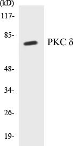
Description
| Product Name: | PKC delta (Phospho-Tyr64) Colorimetric Cell-Based ELISA |
| Product Code: | CBCAB01654 |
| ELISA Type: | Cell-Based |
| Target: | PKC delta (Phospho-Tyr64) |
| Reactivity: | Human, Mouse, Rat |
| Dynamic Range: | > 5000 Cells |
| Detection Method: | Colorimetric 450 nm |
| Format: | 2 x 96-Well Microplates |
The PKC delta (Phospho-Tyr64) Colorimetric Cell-Based ELISA Kit is a convenient, lysate-free, high throughput and sensitive assay kit that can detect PKC delta PKCD protein phosphorylation and expression profile in cells. The kit can be used for measuring the relative amounts of phosphorylated PKC delta PKCD in cultured cells as well as screening for the effects that various treatments, inhibitors (ie. siRNA or chemicals), or activators have on PKC delta PKCD phosphorylation.
Qualitative determination of PKC delta (Phospho-Tyr64) concentration is achieved by an indirect ELISA format. In essence, PKC delta (Phospho-Tyr64) is captured by PKC delta (Phospho-Tyr64)-specific primary (1ø) antibodies while the HRP-conjugated secondary (2ø) antibodies bind the Fc region of the 1ø antibody. Through this binding, the HRP enzyme conjugated to the 2ø antibody can catalyze a colorimetric reaction upon substrate addition. Due to the qualitative nature of the Cell-Based ELISA, multiple normalization methods are needed:
| 1. | A monoclonal antibody specific for human GAPDH is included to serve as an internal positive control in normalizing the target absorbance values. |
| 2. | Following the colorimetric measurement of HRP activity via substrate addition, the Crystal Violet whole-cell staining method may be used to determine cell density. After staining, the results can be analysed by normalizing the absorbance values to cell amounts, by which the plating difference can be adjusted. |
| Database Information: | Gene ID: 5580, UniProt ID: Q05655, OMIM: 176977, Unigene: Hs.155342 |
| Gene Symbol: | PRKCD |
| Sub Type: | Phospho |
| UniProt Protein Function: | Calcium-independent, phospholipid- and diacylglycerol (DAG)-dependent serine/threonine-protein kinase that plays contrasting roles in cell death and cell survival by functioning as a pro-apoptotic protein during DNA damage-induced apoptosis, but acting as an anti-apoptotic protein during cytokine receptor-initiated cell death, is involved in tumor suppression as well as survival of several cancers, is required for oxygen radical production by NADPH oxidase and acts as positive or negative regulator in platelet functional responses. Negatively regulates B cell proliferation and also has an important function in self-antigen induced B cell tolerance induction. Upon DNA damage, activates the promoter of the death-promoting transcription factor BCLAF1/Btf to trigger BCLAF1-mediated p53/TP53 gene transcription and apoptosis. In response to oxidative stress, interact with and activate CHUK/IKKA in the nucleus, causing the phosphorylation of p53/TP53. In the case of ER stress or DNA damage-induced apoptosis, can form a complex with the tyrosine-protein kinase ABL1 which trigger apoptosis independently of p53/TP53. In cytosol can trigger apoptosis by activating MAPK11 or MAPK14, inhibiting AKT1 and decreasing the level of X-linked inhibitor of apoptosis protein (XIAP), whereas in nucleus induces apoptosis via the activation of MAPK8 or MAPK9. Upon ionizing radiation treatment, is required for the activation of the apoptosis regulators BAX and BAK, which trigger the mitochondrial cell death pathway. Can phosphorylate MCL1 and target it for degradation which is sufficient to trigger for BAX activation and apoptosis. Is required for the control of cell cycle progression both at G1/S and G2/M phases. Mediates phorbol 12-myristate 13-acetate (PMA)-induced inhibition of cell cycle progression at G1/S phase by up-regulating the CDK inhibitor CDKN1A/p21 and inhibiting the cyclin CCNA2 promoter activity. In response to UV irradiation can phosphorylate CDK1, which is important for the G2/M DNA damage checkpoint activation. Can protect glioma cells from the apoptosis induced by TNFSF10/TRAIL, probably by inducing increased phosphorylation and subsequent activation of AKT1. Is highly expressed in a number of cancer cells and promotes cell survival and resistance against chemotherapeutic drugs by inducing cyclin D1 (CCND1) and hyperphosphorylation of RB1, and via several pro-survival pathways, including NF-kappa-B, AKT1 and MAPK1/3 (ERK1/2). Can also act as tumor suppressor upon mitogenic stimulation with PMA or TPA. In N-formyl-methionyl-leucyl-phenylalanine (fMLP)-treated cells, is required for NCF1 (p47-phox) phosphorylation and activation of NADPH oxidase activity, and regulates TNF-elicited superoxide anion production in neutrophils, by direct phosphorylation and activation of NCF1 or indirectly through MAPK1/3 (ERK1/2) signaling pathways. May also play a role in the regulation of NADPH oxidase activity in eosinophil after stimulation with IL5, leukotriene B4 or PMA. In collagen-induced platelet aggregation, acts a negative regulator of filopodia formation and actin polymerization by interacting with and negatively regulating VASP phosphorylation. Downstream of PAR1, PAR4 and CD36/GP4 receptors, regulates differentially platelet dense granule secretion; acts as a positive regulator in PAR-mediated granule secretion, whereas it negatively regulates CD36/GP4-mediated granule release. Phosphorylates MUC1 in the C-terminal and regulates the interaction between MUC1 and beta-catenin. The catalytic subunit phosphorylates 14-3-3 proteins (YWHAB, YWHAZ and YWHAH) in a sphingosine-dependent fashion. Phosphorylates ELAVL1 in response to angiotensin-2 treatment (PubMed:18285462). |
| NCBI Summary: | Protein kinase C (PKC) is a family of serine- and threonine-specific protein kinases that can be activated by calcium and the second messenger diacylglycerol. PKC family members phosphorylate a wide variety of protein targets and are known to be involved in diverse cellular signaling pathways. PKC family members also serve as major receptors for phorbol esters, a class of tumor promoters. Each member of the PKC family has a specific expression profile and is believed to play distinct roles in cells. The protein encoded by this gene is one of the PKC family members. Studies both in human and mice demonstrate that this kinase is involved in B cell signaling and in the regulation of growth, apoptosis, and differentiation of a variety of cell types. Alternatively spliced transcript variants encoding the same protein have been observed. [provided by RefSeq, Jul 2008] |
| UniProt Code: | Q05655 |
| NCBI GenInfo Identifier: | 205371776 |
| NCBI Gene ID: | 5580 |
| NCBI Accession: | Q05655.2 |
| UniProt Secondary Accession: | Q05655,Q15144, Q86XJ6, B0KZ81, B2R834, |
| UniProt Related Accession: | Q05655 |
| Molecular Weight: | 80,953 Da |
| NCBI Full Name: | Protein kinase C delta type |
| NCBI Synonym Full Names: | protein kinase C delta |
| NCBI Official Symbol: | PRKCD |
| NCBI Official Synonym Symbols: | MAY1; PKCD; ALPS3; CVID9; nPKC-delta |
| NCBI Protein Information: | protein kinase C delta type |
| UniProt Protein Name: | Protein kinase C delta type |
| UniProt Synonym Protein Names: | Tyrosine-protein kinase PRKCD (EC:2.7.10.2); nPKC-delta |
| Protein Family: | Protein kinase |
| UniProt Gene Name: | PRKCD |
| UniProt Entry Name: | KPCD_HUMAN |
| Component | Quantity |
| 96-Well Cell Culture Clear-Bottom Microplate | 2 plates |
| 10X TBS | 24 mL |
| Quenching Buffer | 24 mL |
| Blocking Buffer | 50 mL |
| 15X Wash Buffer | 50 mL |
| Primary Antibody Diluent | 12 mL |
| 100x Anti-Phospho Target Antibody | 60 µL |
| 100x Anti-Target Antibody | 60 µL |
| Anti-GAPDH Antibody | 60 µL |
| HRP-Conjugated Anti-Rabbit IgG Antibody | 12 mL |
| HRP-Conjugated Anti-Mouse IgG Antibody | 12 mL |
| SDS Solution | 12 mL |
| Stop Solution | 24 mL |
| Ready-to-Use Substrate | 12 mL |
| Crystal Violet Solution | 12 mL |
| Adhesive Plate Seals | 2 seals |
The following materials and/or equipment are NOT provided in this kit but are necessary to successfully conduct the experiment:
- Microplate reader able to measure absorbance at 450 nm and/or 595 nm for Crystal Violet Cell Staining (Optional)
- Micropipettes with capability of measuring volumes ranging from 1 µL to 1 ml
- 37% formaldehyde (Sigma Cat# F-8775) or formaldehyde from other sources
- Squirt bottle, manifold dispenser, multichannel pipette reservoir or automated microplate washer
- Graph paper or computer software capable of generating or displaying logarithmic functions
- Absorbent papers or vacuum aspirator
- Test tubes or microfuge tubes capable of storing ≥1 ml
- Poly-L-Lysine (Sigma Cat# P4832 for suspension cells)
- Orbital shaker (optional)
- Deionized or sterile water
*Note: Protocols are specific to each batch/lot. For the correct instructions please follow the protocol included in your kit.
| Step | Procedure |
| 1. | Seed 200 µL of 20,000 adherent cells in culture medium in each well of a 96-well plate. The plates included in the kit are sterile and treated for cell culture. For suspension cells and loosely attached cells, coat the plates with 100 µL of 10 µg/ml Poly-L-Lysine (not included) to each well of a 96-well plate for 30 minutes at 37 °C prior to adding cells. |
| 2. | Incubate the cells for overnight at 37 °C, 5% CO2. |
| 3. | Treat the cells as desired. |
| 4. | Remove the cell culture medium and rinse with 200 µL of 1x TBS, twice. |
| 5. | Fix the cells by incubating with 100 µL of Fixing Solution for 20 minutes at room temperature. The 4% formaldehyde is used for adherent cells and 8% formaldehyde is used for suspension cells and loosely attached cells. |
| 6. | Remove the Fixing Solution and wash the plate 3 times with 200 µL 1x Wash Buffer for five minutes each time with gentle shaking on the orbital shaker. The plate can be stored at 4 °C for a week. |
| 7. | Add 100 µL of Quenching Buffer and incubate for 20 minutes at room temperature. |
| 8. | Wash the plate 3 times with 1x Wash Buffer for 5 minutes each time. |
| 9. | Add 200 µL of Blocking Buffer and incubate for 1 hour at room temperature. |
| 10. | Wash 3 times with 200 µL of 1x Wash Buffer for 5 minutes each time. |
| 11. | Add 50 µL of 1x primary antibodies Anti-PKC delta (Phospho-Tyr64) Antibody, Anti-PKC delta Antibody and/or Anti-GAPDH Antibody) to the corresponding wells, cover with Parafilm and incubate for 16 hours (overnight) at 4 °C. If the target expression is known to be high, incubate for 2 hours at room temperature. |
| 12. | Wash 3 times with 200 µL of 1x Wash Buffer for 5 minutes each time. |
| 13. | Add 50 µL of 1x secondary antibodies (HRP-Conjugated AntiRabbit IgG Antibody or HRP-Conjugated Anti-Mouse IgG Antibody) to corresponding wells and incubate for 1.5 hours at room temperature. |
| 14. | Wash 3 times with 200 µL of 1x Wash Buffer for 5 minutes each time. |
| 15. | Add 50 µL of Ready-to-Use Substrate to each well and incubate for 30 minutes at room temperature in the dark. |
| 16. | Add 50 µL of Stop Solution to each well and read OD at 450 nm immediately using the microplate reader. |
(Additional Crystal Violet staining may be performed if desired – details of this may be found in the kit technical manual.)


