Description
| Antibody Name: | Phospho-TAK1-S439 Antibody (CAB9437) |
| Antibody SKU: | CAB9437 |
| Antibody Size: | 50µL, 100µL |
| Application: | Western blotting |
| Reactivity: | Mouse |
| Host Species: | Rabbit |
| Immunogen: | A synthesized peptide derived from human Phospho-TAK1 (S439). |
| Application: | Western blotting |
| Recommended Dilution: | WB 1:500 - 1:2000 |
| Reactivity: | Mouse |
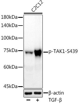
| Positive Samples: | C2C12 |
| Immunogen: | A synthesized peptide derived from human Phospho-TAK1 (S439). |
| Purification Method: | Affinity purification |
| Storage Buffer: | Store at -20°C. Avoid freeze / thaw cycles. Buffer: PBS with 0.02% sodium azide, 50% glycerol, pH7.3. |
| Isotype: | IgG |
| Sequence: | Email for sequence |
| Cellular Location: | Cell membrane, Cytoplasm, Cytoplasmic side, Peripheral membrane protein |
| Calculated MW: | 72kDa |
| Observed MW: | 75KDa |
| Synonyms: | MAP3K7, CSCF, FMD2, MEKK7, TAK1, TGF1a, mitogen-activated protein kinase kinase kinase 7 |
| Background: | The protein encoded by this gene is a member of the serine/threonine protein kinase family. This kinase mediates the signaling transduction induced by TGF beta and morphogenetic protein (BMP), and controls a variety of cell functions including transcription regulation and apoptosis. In response to IL-1, this protein forms a kinase complex including TRAF6, MAP3K7P1/TAB1 and MAP3K7P2/TAB2; this complex is required for the activation of nuclear factor kappa B. This kinase can also activate MAPK8/JNK, MAP2K4/MKK4, and thus plays a role in the cell response to environmental stresses. Four alternatively spliced transcript variants encoding distinct isoforms have been reported. |
 | Western blot analysis of extracts of various cell lines, using at 1:1000 dilution. C2C12 cells were treated by TGF-β (10 ng/ml) at 37℃ for 30 minutes. Secondary antibody: HRP Goat Anti-Rabbit IgG (H+L) at 1:10000 dilution. Lysates/proteins: 25ug per lane. Blocking buffer: 3% nonfat dry milk in TBST. Detection: ECL Basic Kit. Exposure time: 90s. |


