Description
| Antibody Name: | Phospho-SQSTM1/p62-S349 Antibody (CAB20737) |
| Antibody SKU: | CAB20737 |
| Antibody Size: | 50µL, 100µL |
| Application: | Western blotting |
| Reactivity: | Human, Mouse, Rat |
| Host Species: | Rabbit |
| Immunogen: | A synthetic phosphorylated peptide around S349 of human SQSTM1/p62 (NP_003891.1). |
| Application: | Western blotting |
| Recommended Dilution: | WB 1:500 - 1:2000 |
| Reactivity: | Human, Mouse, Rat |
| Positive Samples: | HeLa, C6, NIH/3T3 |
| Immunogen: | A synthetic phosphorylated peptide around S349 of human SQSTM1/p62 (NP_003891.1). |
| Purification Method: | Affinity purification |
| Storage Buffer: | Store at -20°C. Avoid freeze / thaw cycles. Buffer: PBS with 0.02% sodium azide, 50% glycerol, pH7.3. |
| Isotype: | IgG |
| Sequence: | DPST G |
| Cellular Location: | Cytoplasm, Cytoplasmic vesicle, Endoplasmic reticulum, Late endosome, Lysosome, Nucleus, P-body, autophagosome |
| Calculated MW: | 38kDa/47kDa |
| Observed MW: | 62KDa |
| Synonyms: | SQSTM1, A170, DMRV, FTDALS3, NADGP, OSIL, PDB3, ZIP3, p60, p62, p62B |
| Background: | This gene encodes a multifunctional protein that binds ubiquitin and regulates activation of the nuclear factor kappa-B (NF-kB) signaling pathway. The protein functions as a scaffolding/adaptor protein in concert with TNF receptor-associated factor 6 to mediate activation of NF-kB in response to upstream signals. Alternatively spliced transcript variants encoding either the same or different isoforms have been identified for this gene. Mutations in this gene result in sporadic and familial Paget disease of bone. |
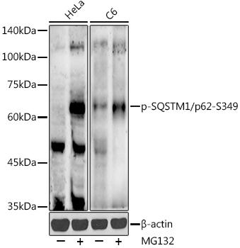
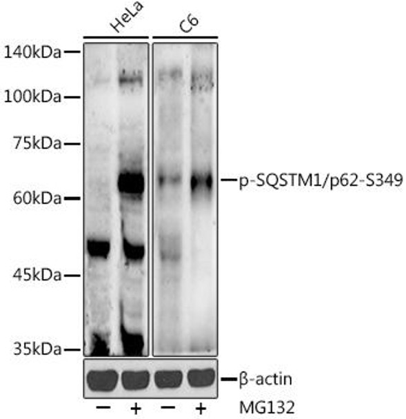
 | Western blot analysis of extracts of various cell lines, using Phospho-SQSTM1/p62-S349 antibody at 1:500 dilution. HeLa cells and C6 cells were treated by MG132(50 μM) at 37℃ for 90 minutes. Secondary antibody: HRP Goat Anti-Rabbit IgG (H+L) at 1:10000 dilution. Lysates/proteins: 25ug per lane. Blocking buffer: 3% nonfat dry milk in TBST. Detection: ECL Enhanced Kit. Exposure time: 90s. |
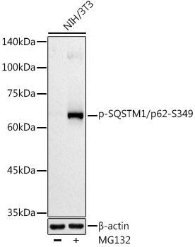
 | Western blot analysis of extracts of NIH/3T3 cells, using Phospho-SQSTM1/p62-S349 antibody at 1:500 dilution. NIH/3T3 cells were treated by MG132(50 μM) at 37℃ for 90 minutes. Secondary antibody: HRP Goat Anti-Rabbit IgG (H+L) at 1:10000 dilution. Lysates/proteins: 25ug per lane. Blocking buffer: 3% nonfat dry milk in TBST. Detection: ECL Basic Kit. Exposure time: 90s. |


