Description
| Antibody Name: | Phospho-ErbB2-Y1139 Antibody (CABP1285) |
| Antibody SKU: | CABP1285 |
| Antibody Size: | 50µL, 100µL |
| Application: | Western blotting |
| Reactivity: | Human |
| Host Species: | Rabbit |
| Immunogen: | A synthesized peptide derived from human Phospho-ErbB2 (Y1139). |
| Application: | Western blotting |
| Recommended Dilution: | WB 1:500 - 1:2000 |
| Reactivity: | Human |
| Positive Samples: | A-431 |
| Immunogen: | A synthesized peptide derived from human Phospho-ErbB2 (Y1139). |
| Purification Method: | Affinity purification |
| Storage Buffer: | Store at -20°C. Avoid freeze / thaw cycles. Buffer: PBS with 0.02% sodium azide, 50% glycerol, pH7.3. |
| Isotype: | IgG |
| Sequence: | Email for sequence |
| Cellular Location: | Cell membrane, Cytoplasm, Nucleus, Nucleus, Single-pass type I membrane protein, perinuclear region |
| Calculated MW: | 180kDa |
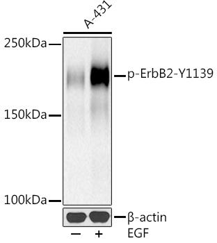
| Observed MW: | 185KDa |
| Synonyms: | ERBB2, CD340, HER-2, HER-2/neu, HER2, MLN 19, NEU, NGL, TKR1, MLN19, erb-b2 receptor tyrosine kinase 2 |
| Background: | This gene encodes a member of the epidermal growth factor (EGF) receptor family of receptor tyrosine kinases. This protein has no ligand binding domain of its own and therefore cannot bind growth factors. However, it does bind tightly to other ligand-bound EGF receptor family members to form a heterodimer, stabilizing ligand binding and enhancing kinase-mediated activation of downstream signalling pathways, such as those involving mitogen-activated protein kinase and phosphatidylinositol-3 kinase. Allelic variations at amino acid positions 654 and 655 of isoform a (positions 624 and 625 of isoform b) have been reported, with the most common allele, Ile654/Ile655, shown here. Amplification and/or overexpression of this gene has been reported in numerous cancers, including breast and ovarian tumors. Alternative splicing results in several additional transcript variants, some encoding different isoforms and others that have not been fully characterized. |
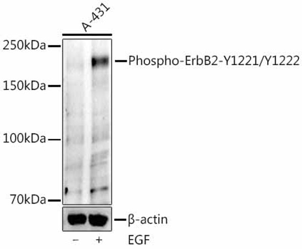
 | Western blot analysis of extracts of A-431 cells, using Phospho-ErbB2-Y1139 at 1:500 dilution. A-431 cells were treated by EGF (100 ng/ml) at 37℃ for 30 minutes after serum-starvation overnight. Secondary antibody: HRP Goat Anti-Rabbit IgG (H+L) at 1:10000 dilution. Lysates/proteins: 25ug per lane. Blocking buffer: 3% nonfat dry milk in TBST. Detection: ECL Basic Kit. Exposure time: 90s. |


