Phospho-BRD4-T204 Antibody (CABP1276)
")
- SKU:
- CABP1276
")
Description
| Antibody Name: | Phospho-BRD4-T204 Antibody (CABP1276) |
| Antibody SKU: | CABP1276 |
| Antibody Size: | 50µL, 100µL |
| Application: | Western blotting |
| Reactivity: | Human, Mouse, Rat |
| Host Species: | Rabbit |
| Immunogen: | A synthetic phosphorylated peptide around T204 of human BRD4 (NP_490597.1). |
| Application: | Western blotting |
| Recommended Dilution: | WB 1:500 - 1:2000 |
| Reactivity: | Human, Mouse, Rat |
| Positive Samples: | HeLa, C6, NIH/3T3 |
| Immunogen: | A synthetic phosphorylated peptide around T204 of human BRD4 (NP_490597.1). |
| Purification Method: | Affinity purification |
| Storage Buffer: | Store at -20°C. Avoid freeze / thaw cycles. Buffer: PBS with 0.02% sodium azide, 50% glycerol, pH7.3. |
| Isotype: | IgG |
| Sequence: | ASTP P |
| Cellular Location: | Chromosome, Nucleus |
| Calculated MW: | 80kDa/88kDa/152kDa |
| Observed MW: | 200KDa |
| Synonyms: | BRD4, CAP, HUNK1, HUNKI, MCAP, bromodomain containing 4 |
| Background: | The protein encoded by this gene is homologous to the murine protein MCAP, which associates with chromosomes during mitosis, and to the human RING3 protein, a serine/threonine kinase. Each of these proteins contains two bromodomains, a conserved sequence motif which may be involved in chromatin targeting. This gene has been implicated as the chromosome 19 target of translocation t(15;19)(q13;p13.1), which defines an upper respiratory tract carcinoma in young people. Two alternatively spliced transcript variants have been described. provided by RefSeq, Jul 2008 |
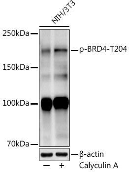 | Western blot analysis of extracts of NIH/3T3 cells, using at 1:1000 dilution. NIH/3T3 cells were treated by Calyculin A (100 nM) for 30 minutes with NaCl (400mM) after serum-starvation 16-20 hours. Secondary antibody: HRP Goat Anti-Rabbit IgG (H+L) at 1:10000 dilution. Lysates/proteins: 25ug per lane. Blocking buffer: 3% nonfat dry milk in TBST. Detection: ECL Enhanced Kit. Exposure time: 90s. |
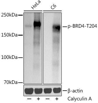 | Western blot analysis of extracts of various cell lines, using at 1:1000 dilution. HeLa cells were treated by Calyculin A (100 nM) at 37℃ for 30 minutes after serum-starvation overnight. C6 cells were treated by Calyculin A (100 nM) at 37℃ for 30 minutes after serum-starvation overnight. Secondary antibody: HRP Goat Anti-Rabbit IgG (H+L) at 1:10000 dilution. Lysates/proteins: 25ug per lane. Blocking buffer: 3% nonfat dry milk in TBST. Detection: ECL Basic Kit. Exposure time: 1s. |
Additional Information
Product Type: |
Antibody |
Antibody Type: |
Monoclonal Antibody |
Reactivity: |
Human |
Reactivity: |
Mouse |
Reactivity: |
Rat |
Host Species: |
Rabbit |
Isotype: |
IgG |
Related Products
")

Phospho-BRD4-S1083 Polyclonal Antibody
Phospho-BRD4-S1083 Polyclonal AntibodyThe product page for this antibody is being prepared. If you have a question please don't hesitate to get in touch!

Phospho-BRD4-T249 Polyclonal Antibody
Phospho-BRD4-T249 Polyclonal AntibodyThe product page for this antibody is being prepared. If you have a question please don't hesitate to get in touch!
")
Phospho-BRD4-S1083 Antibody (CABP1235)
OverviewAntibody Name:Phospho-BRD4-S1083 Antibody (CABP1235)Antibody SKU:CABP1235Antibody Size:50µL, 100µLApplication:Western blottingR
")
Anti-Phospho-BRD4-T240 Antibody (CABP1180)
OverviewProduct Name:Phospho-BRD4-T240 Rabbit pAbProduct Code:CABP1180Size:20uL, 50uL, 100uLSynonyms:BRD4, CAP, HUNK1, HUNKI, MCAP, brom