Description
| Product Name: | p63 (Phospho-Ser455) Colorimetric Cell-Based ELISA |
| Product Code: | CBCAB01351 |
| ELISA Type: | Cell-Based |
| Target: | p63 (Phospho-Ser455) |
| Reactivity: | Human |
| Dynamic Range: | > 5000 Cells |
| Detection Method: | Colorimetric 450 nm |
| Format: | 2 x 96-Well Microplates |
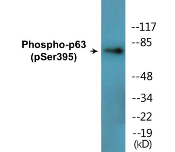
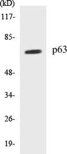
The p63 (Phospho-Ser455) Colorimetric Cell-Based ELISA Kit is a convenient, lysate-free, high throughput and sensitive assay kit that can detect p63 protein phosphorylation and expression profile in cells. The kit can be used for measuring the relative amounts of phosphorylated p63 in cultured cells as well as screening for the effects that various treatments, inhibitors (ie. siRNA or chemicals), or activators have on p63 phosphorylation.
Qualitative determination of p63 (Phospho-Ser455) concentration is achieved by an indirect ELISA format. In essence, p63 (Phospho-Ser455) is captured by p63 (Phospho-Ser455)-specific primary (1ø) antibodies while the HRP-conjugated secondary (2ø) antibodies bind the Fc region of the 1ø antibody. Through this binding, the HRP enzyme conjugated to the 2ø antibody can catalyze a colorimetric reaction upon substrate addition. Due to the qualitative nature of the Cell-Based ELISA, multiple normalization methods are needed:
| 1. | A monoclonal antibody specific for human GAPDH is included to serve as an internal positive control in normalizing the target absorbance values. |
| 2. | Following the colorimetric measurement of HRP activity via substrate addition, the Crystal Violet whole-cell staining method may be used to determine cell density. After staining, the results can be analysed by normalizing the absorbance values to cell amounts, by which the plating difference can be adjusted. |
| Database Information: | Gene ID: 8626, UniProt ID: Q9H3D4, OMIM: 103285/106260/129400/603273/603543/604292/605289, Unigene: Hs.137569 |
| Gene Symbol: | TP63 |
| Sub Type: | Phospho |
| UniProt Protein Function: | Function: Acts as a sequence specific DNA binding transcriptional activator or repressor. The isoforms contain a varying set of transactivation and auto-regulating transactivation inhibiting domains thus showing an isoform specificactivity. Isoform 2 activates RIPK4 transcription. May be required in conjunction with TP73/p73 for initiation of p53/TP53 dependent apoptosis in response to genotoxic insults and the presence of activated oncogenes. Involved in Notch signaling by probably inducing JAG1 and JAG2. Plays a role in the regulation of epithelial morphogenesis. The ratio of DeltaN-type and TA*-type isoforms may govern the maintenance of epithelial stem cell compartments and regulate the initiation of epithelial stratification from the undifferentiated embryonal ectoderm. Required for limb formation from the apical ectodermal ridge. Activates transcription of the p21 promoter. Ref.3 Ref.15 Ref.17 Ref.18 Ref.19 Ref.23 Ref.24 |
| UniProt Protein Details: | Cofactor: Binds 1 zinc ion per subunit By similarity. Subunit structure: Binds DNA as a homotetramer. Isoform compositionof the tetramer may determine transactivation activity. Isoforms Alpha and Gamma interact with HIPK2. Interacts with SSRP1, leading to stimulate coactivator activity. Isoform 1 and isoform 2 interact with WWP1. Interacts with PDS5A. Isoform 5 (via activation domain) interacts with NOC2L. Ref.10 Ref.13 Ref.16 Ref.19 Ref.21 Ref.22 Ref.23 Subcellular location: Nucleus Ref.17 Ref.23. Tissue specificity: Widely expressed, notably in heart, kidney, placenta, prostate, skeletal muscle, testis and thymus, although the precise isoform variesaccording to tissue type. Progenitor cell layers of skin, breast, eye and prostate express high levels of DeltaN-type isoforms. Isoform 10 is predominantly expressed in skin squamous cell carcinomas, but not in normal skin tissues. Ref.3 Ref.8 Ref.14 Domain: The transactivation inhibitory domain (TID) can interact with, and inhibit the activity of the N-terminal transcriptional activation domain of TA*-type isoforms. Ref.17 Ref.18 Post-translational modification: May be sumoylated By similarity.Ubiquitinated. Polyubiquitination involves WWP1 and leads to proteasomal degradation of this protein. Ref.21 Involvement in Disease: Acro-dermato-ungual-lacrimal-tooth syndrome (ADULT syndrome) [MIM:103285]: A form of ectodermal dysplasia. Ectodermal dysplasia defines a heterogeneous group of disorders due to abnormal development of two or more ectodermal structures. ADULT syndrome involves ectrodactyly, syndactyly, finger- and toenail dysplasia, hypoplastic breasts and nipples, intensive freckling, lacrimal duct atresia, frontal alopecia, primary hypodontia and loss of permanent teeth. ADULT syndrome differs significantly from EEC3 syndrome by the absence of facial clefting.Note: The disease is caused by mutations affecting the gene represented in this entry.Ankyloblepharon-ectodermal defects-cleft lip/palate (AEC) [MIM:106260]: An autosomal dominant condition characterized by congenital ectodermal dysplasia with coarse, wiry, sparse hair, dystrophic nails, slight hypohidrosis, scalp infections, ankyloblepharon filiform adnatum, maxillary hypoplasia, hypodontia and cleft lip/palate.Note: The disease is caused by mutations affecting the gene represented in this entry. Ref.29Ectrodactyly, ectodermal dysplasia, and cleft lip/palate syndrome 3 (EEC3) [MIM:604292]: A form of ectodermal dysplasia, a heterogeneous group of disorders due to abnormal development of two or more ectodermal structures. It is an autosomal dominant syndrome characterized by ectrodactyly of hands and feet, ectodermal dysplasia and facial clefting.Note: The disease is caused by mutations affecting the gene represented in this entry. Ref.26 Ref.27 Ref.30 Ref.32Split-hand/foot malformation 4 (SHFM4) [MIM:605289]: A limb malformation involving the central rays of the autopod and presenting with syndactyly, median clefts of the hands and feet, and aplasia and/or hypoplasia of the phalanges, metacarpals, and metatarsals. Some patients have been found to have mental retardation, ectodermal and craniofacial findings, and orofacial clefting.Note: The disease is caused by mutations affecting the gene represented in this entry. Ref.27 Ref.30Limb-mammary syndrome (LMS) [MIM:603543]: Characterized by ectrodactyly, cleft palate and mammary-gland abnormalities.Note: The disease is caused by mutations affecting the gene represented in this entry. Ref.30Defects in TP63 are a cause of cervical, colon, head and neck, lung and ovarian cancers.Ectodermal dysplasia, Rapp-Hodgkin type (EDRH) [MIM:129400]: A form of ectodermal dysplasia, a heterogeneous group of disorders due to abnormal development of two or more ectodermal structures. Characterized by the combination of anhidrotic ectodermal dysplasia, cleft lip, and cleft palate. The clinical syndrome is comprised of a characteristic facies (narrow nose and small mouth), wiry, slow-growing, and uncombable hair, sparse eyelashes and eyebrows, obstructed lacrimal puncta/epiphora, bilateral stenosis of external auditory canals, microsomia, hypodontia, cone-shaped incisors, enamel hypoplasia, dystrophic nails, and cleft lip/cleft palate.Note: The disease is caused by mutations affecting the gene represented in this entry. Ref.33 Ref.34 Ref.35 Ref.36Non-syndromic orofacial cleft 8 (OFC8) [MIM:129400]: A birth defect consisting of cleft lips with or without cleft palate. Cleft lips are associated with cleft palate in two-third of cases. A cleft lip can occur on one or both sides and range in severity from a simple notch in the upper lip to a complete opening in the lip extending into the floor of the nostril and involving the upper gum.Note: The disease is caused by mutations affecting the gene represented in this entry. Sequence similarities: Belongs to the p53 family.Contains 1 SAM (sterile alpha motif) domain. Sequence caution: The sequence AAF43486.1 differs from that shown. Reason: Erroneous initiation. The sequence AAF43487.1 differs from that shown. Reason: Erroneous initiation. The sequence AAF43488.1 differs from that shown. Reason: Erroneous initiation. The sequence AAF43489.1 differs from that shown. Reason: Erroneous initiation. The sequence AAF61624.1 differs from that shown. Reason: Frameshift at position 26. The sequence BAA32592.1 differs from that shown. Reason: Frameshift at position 26. The sequence BAA32593.1 differs from that shown. Reason: Frameshift at position 26. |
| NCBI Summary: | This gene encodes a member of the p53 family of transcription factors. An animal model, p63 -/- mice, has been useful in defining the role this protein plays in the development and maintenance of stratified epithelial tissues. p63 -/- mice have several developmental defects which include the lack of limbs and other tissues, such as teeth and mammary glands, which develop as a result of interactions between mesenchyme and epithelium. Mutations in this gene are associated with ectodermal dysplasia, and cleft lip/palate syndrome 3 (EEC3); split-hand/foot malformation 4 (SHFM4); ankyloblepharon-ectodermal defects-cleft lip/palate; ADULT syndrome (acro-dermato-ungual-lacrimal-tooth); limb-mammary syndrome; Rap-Hodgkin syndrome (RHS); and orofacial cleft 8. Both alternative splicing and the use of alternative promoters results in multiple transcript variants encoding different proteins. Many transcripts encoding different proteins have been reported but the biological validity and the full-length nature of these variants have not been determined. [provided by RefSeq, Jul 2008] |
| UniProt Code: | Q9H3D4 |
| NCBI GenInfo Identifier: | 57013009 |
| NCBI Gene ID: | 8626 |
| NCBI Accession: | Q9H3D4.1 |
| UniProt Secondary Accession: | Q9H3D4,O75080, O75195, O75922, O76078, Q6VEG2, Q6VEG3 Q6VEG4, Q6VFJ1, Q6VFJ2, Q6VFJ3, Q6VH20, |
| UniProt Related Accession: | Q9H3D4 |
| Molecular Weight: | |
| NCBI Full Name: | Tumor protein 63 |
| NCBI Synonym Full Names: | tumor protein p63 |
| NCBI Official Symbol: | TP63 |
| NCBI Official Synonym Symbols: | AIS; KET; LMS; NBP; RHS; p40; p51; p63; EEC3; OFC8; p73H; p73L; SHFM4; TP53L; TP73L; p53CP; TP53CP; B(p51A); B(p51B) |
| NCBI Protein Information: | tumor protein 63; CUSP; transformation-related protein 63; tumor protein p53-competing protein; amplified in squamous cell carcinoma; chronic ulcerative stomatitis protein; keratinocyte transcription factor KET; tumor protein p63 deltaN isoform delta |
| UniProt Protein Name: | Tumor protein 63 |
| UniProt Synonym Protein Names: | Chronic ulcerative stomatitis protein; CUSP; Keratinocyte transcription factor KET; Transformation-related protein 63; TP63; Tumor protein p73-like; p73L; p40; p51 |
| Protein Family: | |
| UniProt Gene Name: | TP63 |
| UniProt Entry Name: | P63_HUMAN |
| Component | Quantity |
| 96-Well Cell Culture Clear-Bottom Microplate | 2 plates |
| 10X TBS | 24 mL |
| Quenching Buffer | 24 mL |
| Blocking Buffer | 50 mL |
| 15X Wash Buffer | 50 mL |
| Primary Antibody Diluent | 12 mL |
| 100x Anti-Phospho Target Antibody | 60 µL |
| 100x Anti-Target Antibody | 60 µL |
| Anti-GAPDH Antibody | 60 µL |
| HRP-Conjugated Anti-Rabbit IgG Antibody | 12 mL |
| HRP-Conjugated Anti-Mouse IgG Antibody | 12 mL |
| SDS Solution | 12 mL |
| Stop Solution | 24 mL |
| Ready-to-Use Substrate | 12 mL |
| Crystal Violet Solution | 12 mL |
| Adhesive Plate Seals | 2 seals |
The following materials and/or equipment are NOT provided in this kit but are necessary to successfully conduct the experiment:
- Microplate reader able to measure absorbance at 450 nm and/or 595 nm for Crystal Violet Cell Staining (Optional)
- Micropipettes with capability of measuring volumes ranging from 1 µL to 1 ml
- 37% formaldehyde (Sigma Cat# F-8775) or formaldehyde from other sources
- Squirt bottle, manifold dispenser, multichannel pipette reservoir or automated microplate washer
- Graph paper or computer software capable of generating or displaying logarithmic functions
- Absorbent papers or vacuum aspirator
- Test tubes or microfuge tubes capable of storing ≥1 ml
- Poly-L-Lysine (Sigma Cat# P4832 for suspension cells)
- Orbital shaker (optional)
- Deionized or sterile water
*Note: Protocols are specific to each batch/lot. For the correct instructions please follow the protocol included in your kit.
| Step | Procedure |
| 1. | Seed 200 µL of 20,000 adherent cells in culture medium in each well of a 96-well plate. The plates included in the kit are sterile and treated for cell culture. For suspension cells and loosely attached cells, coat the plates with 100 µL of 10 µg/ml Poly-L-Lysine (not included) to each well of a 96-well plate for 30 minutes at 37 °C prior to adding cells. |
| 2. | Incubate the cells for overnight at 37 °C, 5% CO2. |
| 3. | Treat the cells as desired. |
| 4. | Remove the cell culture medium and rinse with 200 µL of 1x TBS, twice. |
| 5. | Fix the cells by incubating with 100 µL of Fixing Solution for 20 minutes at room temperature. The 4% formaldehyde is used for adherent cells and 8% formaldehyde is used for suspension cells and loosely attached cells. |
| 6. | Remove the Fixing Solution and wash the plate 3 times with 200 µL 1x Wash Buffer for five minutes each time with gentle shaking on the orbital shaker. The plate can be stored at 4 °C for a week. |
| 7. | Add 100 µL of Quenching Buffer and incubate for 20 minutes at room temperature. |
| 8. | Wash the plate 3 times with 1x Wash Buffer for 5 minutes each time. |
| 9. | Add 200 µL of Blocking Buffer and incubate for 1 hour at room temperature. |
| 10. | Wash 3 times with 200 µL of 1x Wash Buffer for 5 minutes each time. |
| 11. | Add 50 µL of 1x primary antibodies Anti-p63 (Phospho-Ser455) Antibody, Anti-p63 Antibody and/or Anti-GAPDH Antibody) to the corresponding wells, cover with Parafilm and incubate for 16 hours (overnight) at 4 °C. If the target expression is known to be high, incubate for 2 hours at room temperature. |
| 12. | Wash 3 times with 200 µL of 1x Wash Buffer for 5 minutes each time. |
| 13. | Add 50 µL of 1x secondary antibodies (HRP-Conjugated AntiRabbit IgG Antibody or HRP-Conjugated Anti-Mouse IgG Antibody) to corresponding wells and incubate for 1.5 hours at room temperature. |
| 14. | Wash 3 times with 200 µL of 1x Wash Buffer for 5 minutes each time. |
| 15. | Add 50 µL of Ready-to-Use Substrate to each well and incubate for 30 minutes at room temperature in the dark. |
| 16. | Add 50 µL of Stop Solution to each well and read OD at 450 nm immediately using the microplate reader. |
(Additional Crystal Violet staining may be performed if desired – details of this may be found in the kit technical manual.)


