Human Immunology ELISA Kits 6
Human Mothers against decapentaplegic homolog 7 (SMAD7) ELISA Kit
- SKU:
- HUEB0780
- Product Type:
- ELISA Kit
- Size:
- 96 Assays
- Uniprot:
- O15105
- Range:
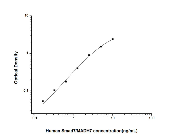
- 0.156-10 ng/mL
- ELISA Type:
- Sandwich
- Synonyms:
- Smad7, MADH7, Mothers Against Decapentaplegic Homolog 7
- Reactivity:
- Human
Description
| Product Name: | Human Mothers against decapentaplegic homolog 7 (SMAD7) ELISA Kit |
| Product Code: | HUEB0780 |
| Alias: | Mothers against decapentaplegic homolog 7, MAD homolog 7, Mothers against DPP homolog 7, Mothers against decapentaplegic homolog 8, MAD homolog 8, Mothers against DPP homolog 8, SMAD family member 7, SMAD 7, Smad7, hSMAD7, SMAD7, MADH7, MADH8 |
| Uniprot: | O15105 |
| Reactivity: | Human |
| Range: | 0.156-10 ng/mL |
| Detection Method: | Sandwich |
| Size: | 96 Assay |
| Storage: | Please see kit components below for exact storage details |
| Note: | For research use only |
| UniProt Protein Function: | SMAD7: Antagonist of signaling by TGF-beta (transforming growth factor) type 1 receptor superfamily members; has been shown to inhibit TGF-beta (Transforming growth factor) and activin signaling by associating with their receptors thus preventing SMAD2 access. Functions as an adapter to recruit SMURF2 to the TGF-beta receptor complex. Also acts by recruiting the PPP1R15A- PP1 complex to TGFBR1, which promotes its dephosphorylation. Positively regulates PDPK1 kinase activity by stimulating its dissociation from the 14-3-3 protein YWHAQ which acts as a negative regulator. Genetic variations in SMAD7 influence susceptibility to colorectal cancer type 3 (CRCS3). Colorectal cancer consists of tumors or cancer of either the colon or rectum or both. Cancers of the large intestine are the second most common form of cancer found in males and females. Symptoms include rectal bleeding, occult blood in stools, bowel obstruction and weight loss. Treatment is based largely on the extent of cancer penetration into the intestinal wall. Surgical cures are possible if the malignancy is confined to the intestine. Risk can be reduced when following a diet which is low in fat and high in fiber. Belongs to the dwarfin/SMAD family. |
| UniProt Protein Details: | Protein type:Transcription factor; DNA-binding Chromosomal Location of Human Ortholog: 18q21.1 Cellular Component: nucleoplasm; transcription factor complex; protein complex; cell-cell adherens junction; cytoplasm; plasma membrane; catenin complex; cytosol; nucleus Molecular Function:collagen binding; protein binding; metal ion binding; ubiquitin protein ligase binding; beta-catenin binding; activin binding; transforming growth factor beta receptor, inhibitory cytoplasmic mediator activity; transcription factor activity Biological Process: transcription initiation from RNA polymerase II promoter; protein stabilization; transcription, DNA-dependent; negative regulation of transcription factor activity; negative regulation of peptidyl-serine phosphorylation; negative regulation of transcription from RNA polymerase II promoter; regulation of activin receptor signaling pathway; negative regulation of BMP signaling pathway; BMP signaling pathway; regulation of transforming growth factor beta receptor signaling pathway; ureteric bud development; positive regulation of protein ubiquitination; positive regulation of proteasomal ubiquitin-dependent protein catabolic process; transforming growth factor beta receptor signaling pathway; positive regulation of cell-cell adhesion; negative regulation of ubiquitin-protein ligase activity; artery morphogenesis; ventricular cardiac muscle morphogenesis; positive regulation of transcription from RNA polymerase II promoter; negative regulation of protein ubiquitination; gene expression; negative regulation of transforming growth factor beta receptor signaling pathway; negative regulation of cell migration Disease: Colorectal Cancer, Susceptibility To, 3 |
| NCBI Summary: | The protein encoded by this gene is a nuclear protein that binds the E3 ubiquitin ligase SMURF2. Upon binding, this complex translocates to the cytoplasm, where it interacts with TGF-beta receptor type-1 (TGFBR1), leading to the degradation of both the encoded protein and TGFBR1. Expression of this gene is induced by TGFBR1. Variations in this gene are a cause of susceptibility to colorectal cancer type 3 (CRCS3). Several transcript variants encoding different isoforms have been found for this gene. [provided by RefSeq] |
| UniProt Code: | O15105 |
| NCBI GenInfo Identifier: | 18418630 |
| NCBI Gene ID: | 4092 |
| NCBI Accession: | |
| UniProt Secondary Accession: | O15105,O14740, Q6DK23, |
| UniProt Related Accession: | O15105 |
| Molecular Weight: | 46,426 Da |
| NCBI Full Name: | Smad7 |
| NCBI Synonym Full Names: | SMAD family member 7 |
| NCBI Official Symbol: | SMAD7 |
| NCBI Official Synonym Symbols: | CRCS3; MADH7; MADH8; FLJ16482 |
| NCBI Protein Information: | mothers against decapentaplegic homolog 7; hSMAD7; MAD homolog 8; OTTHUMP00000163490; mothers against DPP homolog 8; SMAD, mothers against DPP homolog 7; Mothers against decapentaplegic, drosophila, homolog of, 7; MAD (mothers against decapentaplegic, Drosophila) homolog 7 |
| UniProt Protein Name: | Mothers against decapentaplegic homolog 7 |
| UniProt Synonym Protein Names: | Mothers against decapentaplegic homolog 8; MAD homolog 8; Mothers against DPP homolog 8; SMAD family member 7 |
| Protein Family: | Mothers against decapentaplegic |
| UniProt Gene Name: | SMAD7 |
| UniProt Entry Name: | SMAD7_HUMAN |
| Component | Quantity (96 Assays) | Storage |
| ELISA Microplate (Dismountable) | 8×12 strips | -20°C |
| Lyophilized Standard | 2 | -20°C |
| Sample Diluent | 20ml | -20°C |
| Assay Diluent A | 10mL | -20°C |
| Assay Diluent B | 10mL | -20°C |
| Detection Reagent A | 120µL | -20°C |
| Detection Reagent B | 120µL | -20°C |
| Wash Buffer | 30mL | 4°C |
| Substrate | 10mL | 4°C |
| Stop Solution | 10mL | 4°C |
| Plate Sealer | 5 | - |
Other materials and equipment required:
- Microplate reader with 450 nm wavelength filter
- Multichannel Pipette, Pipette, microcentrifuge tubes and disposable pipette tips
- Incubator
- Deionized or distilled water
- Absorbent paper
- Buffer resevoir
*Note: The below protocol is a sample protocol. Protocols are specific to each batch/lot. For the correct instructions please follow the protocol included in your kit.
Allow all reagents to reach room temperature (Please do not dissolve the reagents at 37°C directly). All the reagents should be mixed thoroughly by gently swirling before pipetting. Avoid foaming. Keep appropriate numbers of strips for 1 experiment and remove extra strips from microtiter plate. Removed strips should be resealed and stored at -20°C until the kits expiry date. Prepare all reagents, working standards and samples as directed in the previous sections. Please predict the concentration before assaying. If values for these are not within the range of the standard curve, users must determine the optimal sample dilutions for their experiments. We recommend running all samples in duplicate.
| Step | |
| 1. | Add Sample: Add 100µL of Standard, Blank, or Sample per well. The blank well is added with Sample diluent. Solutions are added to the bottom of micro ELISA plate well, avoid inside wall touching and foaming as possible. Mix it gently. Cover the plate with sealer we provided. Incubate for 120 minutes at 37°C. |
| 2. | Remove the liquid from each well, don't wash. Add 100µL of Detection Reagent A working solution to each well. Cover with the Plate sealer. Gently tap the plate to ensure thorough mixing. Incubate for 1 hour at 37°C. Note: if Detection Reagent A appears cloudy warm to room temperature until solution is uniform. |
| 3. | Aspirate each well and wash, repeating the process three times. Wash by filling each well with Wash Buffer (approximately 400µL) (a squirt bottle, multi-channel pipette,manifold dispenser or automated washer are needed). Complete removal of liquid at each step is essential. After the last wash, completely remove remaining Wash Buffer by aspirating or decanting. Invert the plate and pat it against thick clean absorbent paper. |
| 4. | Add 100µL of Detection Reagent B working solution to each well. Cover with the Plate sealer. Incubate for 60 minutes at 37°C. |
| 5. | Repeat the wash process for five times as conducted in step 3. |
| 6. | Add 90µL of Substrate Solution to each well. Cover with a new Plate sealer and incubate for 10-20 minutes at 37°C. Protect the plate from light. The reaction time can be shortened or extended according to the actual color change, but this should not exceed more than 30 minutes. When apparent gradient appears in standard wells, user should terminatethe reaction. |
| 7. | Add 50µL of Stop Solution to each well. If color change does not appear uniform, gently tap the plate to ensure thorough mixing. |
| 8. | Determine the optical density (OD value) of each well at once, using a micro-plate reader set to 450 nm. User should open the micro-plate reader in advance, preheat the instrument, and set the testing parameters. |
| 9. | After experiment, store all reagents according to the specified storage temperature respectively until their expiry. |
When carrying out an ELISA assay it is important to prepare your samples in order to achieve the best possible results. Below we have a list of procedures for the preparation of samples for different sample types.
| Sample Type | Protocol |
| Serum | If using serum separator tubes, allow samples to clot for 30 minutes at room temperature. Centrifuge for 10 minutes at 1,000x g. Collect the serum fraction and assay promptly or aliquot and store the samples at -80°C. Avoid multiple freeze-thaw cycles. If serum separator tubes are not being used, allow samples to clot overnight at 2-8°C. Centrifuge for 10 minutes at 1,000x g. Remove serum and assay promptly or aliquot and store the samples at -80°C. Avoid multiple freeze-thaw cycles. |
| Plasma | Collect plasma using EDTA or heparin as an anticoagulant. Centrifuge samples at 4°C for 15 mins at 1000 × g within 30 mins of collection. Collect the plasma fraction and assay promptly or aliquot and store the samples at -80°C. Avoid multiple freeze-thaw cycles. Note: Over haemolysed samples are not suitable for use with this kit. |
| Urine & Cerebrospinal Fluid | Collect the urine (mid-stream) in a sterile container, centrifuge for 20 mins at 2000-3000 rpm. Remove supernatant and assay immediately. If any precipitation is detected, repeat the centrifugation step. A similar protocol can be used for cerebrospinal fluid. |
| Cell culture supernatant | Collect the cell culture media by pipette, followed by centrifugation at 4°C for 20 mins at 1500 rpm. Collect the clear supernatant and assay immediately. |
| Cell lysates | Solubilize cells in lysis buffer and allow to sit on ice for 30 minutes. Centrifuge tubes at 14,000 x g for 5 minutes to remove insoluble material. Aliquot the supernatant into a new tube and discard the remaining whole cell extract. Quantify total protein concentration using a total protein assay. Assay immediately or aliquot and store at ≤ -20 °C. |
| Tissue homogenates | The preparation of tissue homogenates will vary depending upon tissue type. Rinse tissue with 1X PBS to remove excess blood & homogenize in 20ml of 1X PBS (including protease inhibitors) and store overnight at ≤ -20°C. Two freeze-thaw cycles are required to break the cell membranes. To further disrupt the cell membranes you can sonicate the samples. Centrifuge homogenates for 5 mins at 5000xg. Remove the supernatant and assay immediately or aliquot and store at -20°C or -80°C. |
| Tissue lysates | Rinse tissue with PBS, cut into 1-2 mm pieces, and homogenize with a tissue homogenizer in PBS. Add an equal volume of RIPA buffer containing protease inhibitors and lyse tissues at room temperature for 30 minutes with gentle agitation. Centrifuge to remove debris. Quantify total protein concentration using a total protein assay. Assay immediately or aliquot and store at ≤ -20 °C. |
| Breast Milk | Collect milk samples and centrifuge at 10,000 x g for 60 min at 4°C. Aliquot the supernatant and assay. For long term use, store samples at -80°C. Minimize freeze/thaw cycles. |


