Human Cell Biology ELISA Kits 3
Human MCP-1 (Monocyte Chemotactic Protein 1) CLIA Kit (HUES00020)
- SKU:
- HUES00020
- Product Type:
- ELISA Kit
- ELISA Type:
- CLIA Kit
- Size:
- 96 Assays
- Sensitivity:
- 4.69pg/mL
- Range:
- 7.81-500pg/mL
- ELISA Type:
- Sandwich
- Synonyms:
- CCL2, GDCF-2, HC11, HSMCR30, MCAF, MCP1, SCYA2, SMC-CF
- Reactivity:
- Human
- Sample Type:
- Serum, plasma and other biological fluids
- Research Area:
- Cell Biology
Description
| Assay type: | Sandwich |
| Format: | 96T |
| Assay time: | 4.5h |
| Reactivity: | Human |
| Detection method: | Chemiluminescence |
| Detection range: | 7.81-500 pg/mL |
| Sensitivity: | 4.69 pg/mL |
| Sample volume: | 100µL |
| Sample type: | Serum, plasma and other biological fluids |
| Repeatability: | CV < 15% |
| Specificity: | This kit recognizes Human MCP-1 in samples. No significant cross-reactivity or interference between Human MCP-1 and analogues was observed. |
This kit uses Sandwich-CLIA as the method. The micro CLIA plate provided in this kit has been pre-coated with an antibody specific to Human MCP-1. Standards or samples are added to the appropriate micro CLIA plate wells and combined with the specific antibody. Then a biotinylated detection antibody specific for Human MCP-1 and Avidin-Horseradish Peroxidase (HRP) conjugate are added to each micro plate well successively and incubated. Free components are washed away. The substrate solution is added to each well. Only those wells that contain Human MCP-1, biotinylated detection antibody and Avidin-HRP conjugate will appear fluorescence. The Relative light unit (RLU) value is measured spectrophotometrically by the Chemiluminescence immunoassay analyzer. The RLU value is positively associated with the concentration of Human MCP-1. The concentration of Human MCP-1 in the samples can be calculated by comparing the RLU of the samples to the standard curve.
| UniProt Protein Function: | CCL2: Chemotactic factor that attracts monocytes and basophils but not neutrophils or eosinophils. Augments monocyte anti-tumor activity. Has been implicated in the pathogenesis of diseases characterized by monocytic infiltrates, like psoriasis, rheumatoid arthritis or atherosclerosis. May be involved in the recruitment of monocytes into the arterial wall during the disease process of atherosclerosis. Monomer or homodimer; in equilibrium. Binds to CCR2 and CCR4. Is tethered on endothelial cells by glycosaminoglycan (GAG) side chains of proteoglycans. Belongs to the intercrine beta (chemokine CC) family. |
| UniProt Protein Details: | Protein type:Secreted; Chemokine; Secreted, signal peptide; Motility/polarity/chemotaxis Chromosomal Location of Human Ortholog: 17q11. 2-q12 Cellular Component: extracellular space; rough endoplasmic reticulum; perinuclear region of cytoplasm; endocytic vesicle; dendrite; extracellular region; synapse; perikaryon; nerve terminal Molecular Function:heparin binding; chemokine activity; CCR2 chemokine receptor binding; receptor binding; protein kinase activity Biological Process: maternal process involved in pregnancy; protein amino acid phosphorylation; response to antibiotic; monocyte chemotaxis; regulation of cell shape; cell surface receptor linked signal transduction; response to vitamin B3; transforming growth factor beta receptor signaling pathway; negative regulation of neuron apoptosis; cellular homeostasis; cell adhesion; neutrophil chemotaxis; organ regeneration; response to amino acid stimulus; JAK-STAT cascade; positive regulation of tumor necrosis factor production; G-protein signaling, coupled to cyclic nucleotide second messenger; organ morphogenesis; unfolded protein response, activation of signaling protein activity; response to ethanol; cellular response to insulin stimulus; response to bacterium; response to heat; response to mechanical stimulus; positive regulation of endothelial cell proliferation; response to activity; response to progesterone stimulus; positive regulation of nitric-oxide synthase biosynthetic process; positive regulation of collagen biosynthetic process; signal transduction; chemotaxis; positive regulation of synaptic transmission; positive regulation of cellular extravasation; protein kinase B signaling cascade; response to wounding; lipopolysaccharide-mediated signaling pathway; response to gamma radiation; angiogenesis; inflammatory response; lymphocyte chemotaxis; aging; unfolded protein response; cytokine and chemokine mediated signaling pathway; MAPKKK cascade; cytoskeleton organization and biogenesis; viral genome replication; macrophage chemotaxis; humoral immune response; leukocyte migration during inflammatory response; cellular calcium ion homeostasis; G-protein coupled receptor protein signaling pathway; negative regulation of angiogenesis; positive regulation of leukocyte mediated cytotoxicity; cellular protein metabolic process; maternal process involved in parturition; response to hypoxia; positive regulation of T cell activation; vascular endothelial growth factor receptor signaling pathway; astrocyte cell migration Disease: Neural Tube Defects; Mycobacterium Tuberculosis, Susceptibility To; Human Immunodeficiency Virus Type 1, Susceptibility To |
| NCBI Summary: | This gene is one of several cytokine genes clustered on the q-arm of chromosome 17. Chemokines are a superfamily of secreted proteins involved in immunoregulatory and inflammatory processes. The superfamily is divided into four subfamilies based on the arrangement of N-terminal cysteine residues of the mature peptide. This chemokine is a member of the CC subfamily which is characterized by two adjacent cysteine residues. This cytokine displays chemotactic activity for monocytes and basophils but not for neutrophils or eosinophils. It has been implicated in the pathogenesis of diseases characterized by monocytic infiltrates, like psoriasis, rheumatoid arthritis and atherosclerosis. It binds to chemokine receptors CCR2 and CCR4. [provided by RefSeq, Jul 2013] |
| UniProt Code: | P13500 |
| NCBI GenInfo Identifier: | 126842 |
| NCBI Gene ID: | 6347 |
| NCBI Accession: | P13500. 1 |
| UniProt Secondary Accession: | P13500,Q9UDF3, B2R4V3, |
| UniProt Related Accession: | P13500 |
| Molecular Weight: | 11,025 Da |
| NCBI Full Name: | C-C motif chemokine 2 |
| NCBI Synonym Full Names: | chemokine (C-C motif) ligand 2 |
| NCBI Official Symbol: | CCL2 |
| NCBI Official Synonym Symbols: | HC11; MCAF; MCP1; MCP-1; SCYA2; GDCF-2; SMC-CF; HSMCR30 |
| NCBI Protein Information: | C-C motif chemokine 2; small-inducible cytokine A2; monocyte secretory protein JE; monocyte chemotactic protein 1; monocyte chemoattractant protein 1; monocyte chemoattractant protein-1; monocyte chemotactic and activating factor; small inducible cytokine subfamily A (Cys-Cys), member 2; small inducible cytokine A2 (monocyte chemotactic protein 1, homologous to mouse Sig-je) |
| UniProt Protein Name: | C-C motif chemokine 2 |
| UniProt Synonym Protein Names: | HC11; Monocyte chemoattractant protein 1; Monocyte chemotactic and activating factor; MCAF; Monocyte chemotactic protein 1; MCP-1; Monocyte secretory protein JE; Small-inducible cytokine A2 |
| Protein Family: | C-C motif chemokine |
| UniProt Gene Name: | CCL2 |
| UniProt Entry Name: | CCL2_HUMAN |
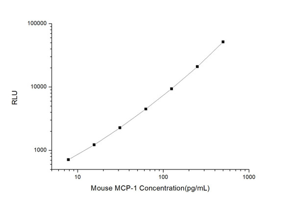
As the RLU values of the standard curve may vary according to the conditions of the actual assay performance (e. g. operator, pipetting technique, washing technique or temperature effects), the operator should establish a standard curve for each test. Typical standard curve and data is provided below for reference only.
| Concentration (pg/mL) | RLU | Average | Corrected |
| 500 | 49786 53288 | 51537 | 51508 |
| 250 | 19868 22128 | 20998 | 20969 |
| 125 | 9781 9011 | 9396 | 9367 |
| 62.5 | 4178 4846 | 4512 | 4483 |
| 31.25 | 2460 2138 | 2299 | 2270 |
| 15.63 | 1256 1244 | 1250 | 1221 |
| 7.81 | 714 766 | 740 | 711 |
| 0 | 28 30 | 29 | -- |
Precision
Intra-assay Precision (Precision within an assay): 3 samples with low, mid range and high level Human MCP-1 were tested 20 times on one plate, respectively.
Inter-assay Precision (Precision between assays): 3 samples with low, mid range and high level Human MCP-1 were tested on 3 different plates, 20 replicates in each plate.
| Intra-assay Precision | Inter-assay Precision | |||||
| Sample | 1 | 2 | 3 | 1 | 2 | 3 |
| n | 20 | 20 | 20 | 20 | 20 | 20 |
| Mean (pg/mL) | 26.97 | 50.59 | 231.90 | 24.71 | 45.93 | 218.62 |
| Standard deviation | 3.42 | 4.44 | 20.06 | 2.94 | 3.69 | 22.06 |
| C V (%) | 12.68 | 8.78 | 8.65 | 11.90 | 8.03 | 10.09 |
Recovery
The recovery of Human MCP-1 spiked at three different levels in samples throughout the range of the assay was evaluated in various matrices.
| Sample Type | Range (%) | Average Recovery (%) |
| Serum (n=5) | 100-112 | 106 |
| EDTA plasma (n=5) | 90-104 | 98 |
| Cell culture media (n=5) | 93-107 | 99 |
Linearity
Samples were spiked with high concentrations of Human MCP-1 and diluted with Reference Standard & Sample Diluent to produce samples with values within the range of the assay.
| Serum (n=5) | EDTA plasma (n=5) | Cell culture media (n=5) | ||
| 1:2 | Range (%) | 96-112 | 88-102 | 88-99 |
| Average (%) | 102 | 93 | 94 | |
| 1:4 | Range (%) | 91-106 | 90-105 | 93-111 |
| Average (%) | 97 | 96 | 101 | |
| 1:8 | Range (%) | 93-106 | 102-115 | 101-114 |
| Average (%) | 99 | 109 | 108 | |
| 1:16 | Range (%) | 84-96 | 96-108 | 97-114 |
| Average (%) | 91 | 102 | 105 |
An unopened kit can be stored at 4°C for 1 month. If the kit is not used within 1 month, store the items separately according to the following conditions once the kit is received.
| Item | Specifications | Storage |
| Micro CLIA Plate(Dismountable) | 8 wells ×12 strips | -20°C, 6 months |
| Reference Standard | 2 vials | |
| Concentrated Biotinylated Detection Ab (100×) | 1 vial, 120 µL | |
| Concentrated HRP Conjugate (100×) | 1 vial, 120 µL | -20°C(shading light), 6 months |
| Reference Standard & Sample Diluent | 1 vial, 20 mL | 4°C, 6 months |
| Biotinylated Detection Ab Diluent | 1 vial, 14 mL | |
| HRP Conjugate Diluent | 1 vial, 14 mL | |
| Concentrated Wash Buffer (25×) | 1 vial, 30 mL | |
| Substrate Reagent A | 1 vial, 5 mL | 4°C (shading light) |
| Substrate Reagent B | 1 vial, 5 mL | 4°C (shading light) |
| Plate Sealer | 5 pieces | |
| Product Description | 1 copy | |
| Certificate of Analysis | 1 copy |
- Set standard, test sample and control (zero) wells on the pre-coated plate and record theirpositions. It is recommended to measure each standard and sample in duplicate. Note: addall solutions to the bottom of the plate wells while avoiding contact with the well walls. Ensuresolutions do not foam when adding to the wells.
- Aliquot 100µl of standard solutions into the standard wells.
- Add 100µl of Sample / Standard dilution buffer into the control (zero) well.
- Add 100µl of properly diluted sample (serum, plasma, tissue homogenates and otherbiological fluids. ) into test sample wells.
- Cover the plate with the sealer provided in the kit and incubate for 90 min at 37°C.
- Aspirate the liquid from each well, do not wash. Immediately add 100µL of BiotinylatedDetection Ab working solution to each well. Cover the plate with a plate seal and gently mix. Incubate for 1 hour at 37°C.
- Aspirate or decant the solution from the plate and add 350µL of wash buffer to each welland incubate for 1-2 minutes at room temperature. Aspirate the solution from each well andclap the plate on absorbent filter paper to dry. Repeat this process 3 times. Note: a microplatewasher can be used in this step and other wash steps.
- Add 100µL of HRP Conjugate working solution to each well. Cover with a plate seal andincubate for 30 min at 37°C.
- Aspirate or decant the solution from each well. Repeat the wash process for five times asconducted in step 7.
- Add 100µL of Substrate mixture solution to each well. Cover with a new plate seal andincubate for no more than 5 min at 37°C. Protect the plate from light.
- Determine the RLU value of each well immediately.


