Description
Human IFN-gamma ELISpot Kit
Assay Genie ELISpot is a highly specific immunoassay for the analysis of IFN-γ production and secretion from T-cells at a single cell level in conditions closely comparable to the in-vivo environment with minimal cell manipulation. This technique is designed to determine the frequency of IFN-γ producing cells under a given stimulation and the comparison of such frequency against a specific treatment or pathological state. Utilising sandwich immuno-enzyme technology, Assay Genie ELISpot assays can detect both secreted IFN-γ (qualitative analysis) and single cells that produce IFN-γ (quantitative analysis). Cell secreted IFN-γ is captured by coated antibodies avoiding diffusion in supernatant, protease degradation or binding on soluble membrane receptors. After cell removal, the captured IFN-γ is revealed by tracer antibodies and appropriate conjugates.
| Product type: | ELISpot Kit |
| Size: | 1 x 96 Assays |
| Target species: | Human |
| Specificity: | Recognizes natural human IFN-γ |
| Incubation: | 3h after cell stimulation |
| Kit content: | Assay Genie Pre-coated ELISpot kits include precoated PVDF plates, Detection antibody, Alkaline phosphatase conjugate, BSA, BCIP/NBT ready-to-use substrate buffer. |
| Synonyms: | IFN-g, IFN-gamma |
| Uniprot: | P01579 |
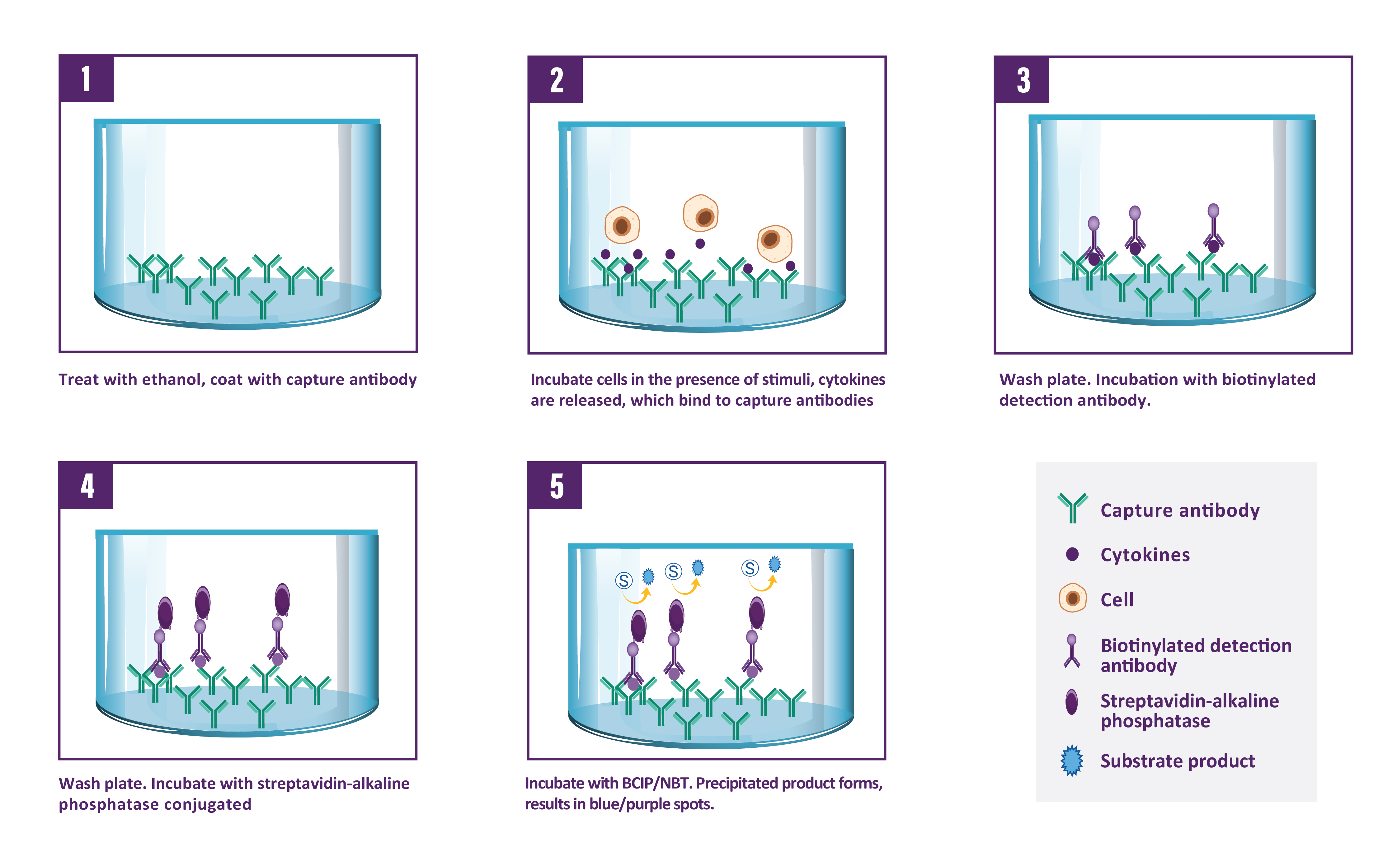
A capture antibody highly specific for IFN-γ is coated to the wells of a PVDF bottomed 96 well microtitre plate either during kit manufacture or in the laboratory. The plate is then blocked to minimise any non-antibody dependent unspecific binding and washed. Cell suspension and stimulant are added and the plate incubated allowing the specific antibodies to bind any IFN-γ produced. Cells are then removed by washing prior to the addition of Biotinylated detection antibodies which bind to the previously captured IFN-γ. Enzyme conjugated streptavidin is then added binding to the detection antibodies. Following incubation and washing, substrate is then applied to the wells resulting in coloured spots which can be quantified using appropriate analysis software or manually using a microscope.

| Step | Procedure |
| 1. | For PVDF membrane activation, add 25 µl of 35% ethanol to every well. |
| 2. | Incubate plate at room temperature (RT) for 30 seconds. |
| 3. | Empty the wells by flicking the plate over a sink & gently tapping on absorbent paper. Thoroughly wash the plate 3x with 100 µl of PBS 1X per well. |
| 4. | Add 100 µl of diluted capture antibody to every well. |
| 5. | Cover the plate and incubate at 4°C overnight. |
| 6. | Empty the wells as previous and wash the plate once with 100 µl of PBS 1X per well. |
| 7. | Add 100 µl of blocking buffer to every well. |
| 8. | Cover the plate and incubate at RT for 2 hours. |
| 9. | Empty the wells as previous and thoroughly wash once with 100 µl of PBS 1X per well. |
| 10. | Add 100 µl of sample, positive and negative controls cell suspension to appropriate wells providing the required concentration of cells and stimulant. |
| 11. | Cover the plate and incubate at 37°C in a CO2 incubator for an appropriate length of time (15-20 hours). Note: do not agitate or move the plate during this incubation. |
| 12. | Empty the wells and remove excess solution then add 100 µl of Wash Buffer to every well. |
| 13. | Incubate the plate at 4°C for 10 min. |
| 14. | Empty the wells as previous and wash the plate 3x with 100 µl of Wash Buffer. |
| 15. | Add 100 µl of diluted detection antibody to every well. |
| 16. | Cover the plate and incubate at RT for 1 hour 30 min. |
| 17. | Empty the wells as previous and wash the plate 3x with 100 µl of Wash Buffer. |
| 18. | Add 100 µl of diluted Streptavidin-conjugate to every well. |
| 19. | Cover the plate and incubate at RT following the supplier's instructions. |
| 20. | Empty the wells and wash the plate 3x with 100 µl of Wash Buffer. |
| 21. | Peel of the plate bottom and wash both sides of the membrane 3x under running distilled water, once washing complete remove any excess solution by repeated tapping on absorbent paper. |
| 22. | Add 100 µl of ready-to-use substrate buffer to every well. |
| 23. | Following the supplier's instructions, incubate the plate for 5-15 min monitoring spot formation visually throughout the incubation period to assess sufficient colour development. |
| 24. | Empty the wells and rinse both sides of the membrane 3x under running distilled water. Completely remove any excess solution by gentle repeated tapping on absorbent paper Read Spots: allow the wells to dry and then read results. The frequency of the resulting coloured spots. |
| UniProt Protein Function: | IFNG: Produced by lymphocytes activated by specific antigens or mitogens. IFN-gamma, in addition to having antiviral activity, has important immunoregulatory functions. It is a potent activator of macrophages, it has antiproliferative effects on transformed cells and it can potentiate the antiviral and antitumor effects of the type I interferons. Homodimer. Released primarily from activated T lymphocytes. Belongs to the type II (or gamma) interferon family. |
| UniProt Protein Details: | Protein type:Membrane protein, integral; Secreted; Secreted, signal peptide; Cytokine Chromosomal Location of Human Ortholog: 12q14 Cellular Component: extracellular space; extracellular region; external side of plasma membrane Molecular Function:interferon-gamma receptor binding; cytokine activity Biological Process: positive regulation of isotype switching to IgG isotypes; positive regulation of nitric oxide biosynthetic process; apoptosis; positive regulation of interleukin-23 production; negative regulation of smooth muscle cell proliferation; positive regulation of interleukin-12 production; positive regulation of osteoclast differentiation; positive regulation of interleukin-6 biosynthetic process; negative regulation of epithelial cell differentiation; positive regulation of interleukin-1 beta secretion; positive regulation of killing of cells of another organism; negative regulation of transcription from RNA polymerase II promoter; sensory perception of mechanical stimulus; positive regulation of membrane protein ectodomain proteolysis; cell surface receptor linked signal transduction; positive regulation of MHC class II biosynthetic process; positive regulation of cell proliferation; positive regulation of mesenchymal cell proliferation; positive regulation of T cell proliferation; cell cycle arrest; defense response to virus; regulation of the force of heart contraction; positive regulation of peptidyl-serine phosphorylation of STAT protein; response to drug; positive regulation of synaptic transmission, cholinergic; neutrophil chemotaxis; adaptive immune response; CD8-positive, alpha-beta T cell differentiation during immune response; negative regulation of myelination; unfolded protein response; response to virus; cytokine and chemokine mediated signaling pathway; defense response to protozoan; positive regulation of tumor necrosis factor production; humoral immune response; antigen processing and presentation; positive regulation of tyrosine phosphorylation of Stat1 protein; positive regulation of chemokine biosynthetic process; negative regulation of interleukin-17 production; positive regulation of interleukin-12 biosynthetic process; protein import into nucleus, translocation; defense response to bacterium; neutrophil apoptosis; positive regulation of transcription from RNA polymerase II promoter; cell motility; positive regulation of neuron differentiation; regulation of insulin secretion Disease: Hepatitis C Virus, Susceptibility To; Aplastic Anemia; Tuberous Sclerosis 2; Mycobacterium Tuberculosis, Susceptibility To; Human Immunodeficiency Virus Type 1, Susceptibility To |
| NCBI Summary: | This gene encodes a member of the type II interferon family. The protein encoded is a soluble cytokine with antiviral, immunoregulatory and anti-tumor properties and is a potent activator of macrophages. Mutations in this gene are associated with aplastic anemia.[provided by RefSeq, Nov 2009] |
| UniProt Code: | P01579 |
| NCBI GenInfo Identifier: | 124479 |
| NCBI Gene ID: | 3458 |
| NCBI Accession: | P01579.1 |
| UniProt Secondary Accession: | P01579,Q53ZV4, B5BU88, |
| UniProt Related Accession: | P01579 |
| Molecular Weight: | |
| NCBI Full Name: | Interferon gamma |
| NCBI Synonym Full Names: | interferon, gamma |
| NCBI Official Symbol: | IFNG‚ ‚ |
| NCBI Official Synonym Symbols: | IFG; IFI‚ ‚ |
| NCBI Protein Information: | interferon gamma; IFN-gamma; immune interferon |
| UniProt Protein Name: | Interferon gamma |
| UniProt Synonym Protein Names: | Immune interferon |
| Protein Family: | Interferon |
| UniProt Gene Name: | IFNG‚ ‚ |
| UniProt Entry Name: | IFNG_HUMAN |


