Human Signal Transduction ELISA Kits
Human HB beta (Hemoglobin Beta) CLIA Kit (HUES01147)
- SKU:
- HUES01147
- Product Type:
- ELISA Kit
- ELISA Type:
- CLIA Kit
- Size:
- 96 Assays
- Sensitivity:
- 0.19Mug/mL
- Range:
- 0.31-20Mug/mL
- ELISA Type:
- Sandwich
- Reactivity:
- Human
- Sample Type:
- Serum, plasma and other biological fluids
- Research Area:
- Signal Transduction
Description
| Assay type: | Sandwich |
| Format: | 96T |
| Assay time: | 4.5h |
| Reactivity: | Human |
| Detection method: | Chemiluminescence |
| Detection range: | 0.31-20 µg/mL |
| Sensitivity: | 0.19 µg/mL |
| Sample volume: | 100µL |
| Sample type: | Serum, plasma and other biological fluids |
| Repeatability: | CV < 15% |
| Specificity: | This kit recognizes Human HB beta in samples. No significant cross-reactivity or interference between Human HB beta and analogues was observed. |
This kit uses Sandwich-CLIA as the method. The micro CLIA plate provided in this kit has been pre-coated with an antibody specific to Human HB beta. Standards or samples are added to the appropriate micro CLIA plate wells and combined with the specific antibody. Then a biotinylated detection antibody specific for Human HB beta and Avidin-Horseradish Peroxidase (HRP) conjugate are added to each micro plate well successively and incubated. Free components are washed away. The substrate solution is added to each well. Only those wells that contain Human HB beta, biotinylated detection antibody and Avidin-HRP conjugate will appear fluorescence. The Relative light unit (RLU) value is measured spectrophotometrically by the Chemiluminescence immunoassay analyzer. The RLU value is positively associated with the concentration of Human HB beta. The concentration of Human HB beta in the samples can be calculated by comparing the RLU of the samples to the standard curve.
| UniProt Protein Function: | HBB: Involved in oxygen transport from the lung to the various peripheral tissues. Defects in HBB may be a cause of Heinz body anemias (HEIBAN). This is a form of non-spherocytic hemolytic anemia of Dacie type 1. After splenectomy, which has little benefit, basophilic inclusions called Heinz bodies are demonstrable in the erythrocytes. Before splenectomy, diffuse or punctate basophilia may be evident. Most of these cases are probably instances of hemoglobinopathy. The hemoglobin demonstrates heat lability. Heinz bodies are observed also with the Ivemark syndrome (asplenia with cardiovascular anomalies) and with glutathione peroxidase deficiency. Defects in HBB are the cause of beta-thalassemia (B-THAL). A form of thalassemia. Thalassemias are common monogenic diseases occurring mostly in Mediterranean and Southeast Asian populations. The hallmark of beta-thalassemia is an imbalance in globin-chain production in the adult HbA molecule. Absence of beta chain causes beta(0)-thalassemia, while reduced amounts of detectable beta globin causes beta(+)-thalassemia. In the severe forms of beta-thalassemia, the excess alpha globin chains accumulate in the developing erythroid precursors in the marrow. Their deposition leads to a vast increase in erythroid apoptosis that in turn causes ineffective erythropoiesis and severe microcytic hypochromic anemia. Clinically, beta-thalassemia is divided into thalassemia major which is transfusion dependent, thalassemia intermedia (of intermediate severity), and thalassemia minor that is asymptomatic. Defects in HBB are the cause of sickle cell anemia (SKCA); also known as sickle cell disease. Sickle cell anemia is characterized by abnormally shaped red cells resulting in chronic anemia and periodic episodes of pain, serious infections and damage to vital organs. Normal red blood cells are round and flexible and flow easily through blood vessels, but in sickle cell anemia, the abnormal hemoglobin (called Hb S) causes red blood cells to become stiff. They are C-shaped and resembles a sickle. These stiffer red blood cells can led to microvascular occlusion thus cutting off the blood supply to nearby tissues. Defects in HBB are the cause of beta-thalassemia dominant inclusion body type (B-THALIB). An autosomal dominant form of beta thalassemia characterized by moderate anemia, lifelong jaundice, cholelithiasis and splenomegaly, marked morphologic changes in the red cells, erythroid hyperplasia of the bone marrow with increased numbers of multinucleate red cell precursors, and the presence of large inclusion bodies in the normoblasts, both in the marrow and in the peripheral blood after splenectomy. Belongs to the globin family. |
| UniProt Protein Details: | Protein type:Carrier Chromosomal Location of Human Ortholog: 11p15. 5 Cellular Component: hemoglobin complex; extracellular region; cytosol Molecular Function:haptoglobin binding; protein binding; peroxidase activity; hemoglobin binding; iron ion binding; heme binding; oxygen binding; oxygen transporter activity Biological Process: receptor-mediated endocytosis; positive regulation of nitric oxide biosynthetic process; response to hydrogen peroxide; nitric oxide transport; bicarbonate transport; hydrogen peroxide catabolic process; oxygen transport; protein heterooligomerization; regulation of blood pressure; regulation of blood vessel size; blood coagulation Disease: Fetal Hemoglobin Quantitative Trait Locus 1; Beta-thalassemia; Sickle Cell Anemia; Heinz Body Anemias; Beta-thalassemia, Dominant Inclusion Body Type; Malaria, Susceptibility To; Alpha-thalassemia |
| NCBI Summary: | The alpha (HBA) and beta (HBB) loci determine the structure of the 2 types of polypeptide chains in adult hemoglobin, Hb A. The normal adult hemoglobin tetramer consists of two alpha chains and two beta chains. Mutant beta globin causes sickle cell anemia. Absence of beta chain causes beta-zero-thalassemia. Reduced amounts of detectable beta globin causes beta-plus-thalassemia. The order of the genes in the beta-globin cluster is 5'-epsilon -- gamma-G -- gamma-A -- delta -- beta--3'. [provided by RefSeq, Jul 2008] |
| UniProt Code: | P68871 |
| NCBI GenInfo Identifier: | 56749856 |
| NCBI Gene ID: | 3043 |
| NCBI Accession: | P68871. 2 |
| UniProt Secondary Accession: | P68871,P02023, Q13852, Q14481, Q14510, Q45KT0, Q549N7 Q6FI08, Q6R7N2, Q8IZI1, A4GX73, B2ZUE0, |
| UniProt Related Accession: | P68871 |
| Molecular Weight: | 15,998 Da |
| NCBI Full Name: | Hemoglobin subunit beta |
| NCBI Synonym Full Names: | hemoglobin, beta |
| NCBI Official Symbol: | HBB |
| NCBI Official Synonym Symbols: | CD113t-C; beta-globin |
| NCBI Protein Information: | hemoglobin subunit beta; beta globin chain; hemoglobin beta chain |
| UniProt Protein Name: | Hemoglobin subunit beta |
| UniProt Synonym Protein Names: | Beta-globin; Hemoglobin beta chainCleaved into the following 2 chains:LVV-hemorphin-7; Spinorphin |
| Protein Family: | Hemoglobin |
| UniProt Gene Name: | HBB |
| UniProt Entry Name: | HBB_HUMAN |
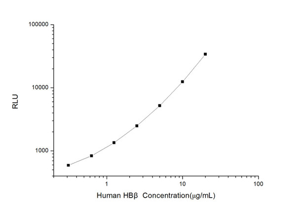
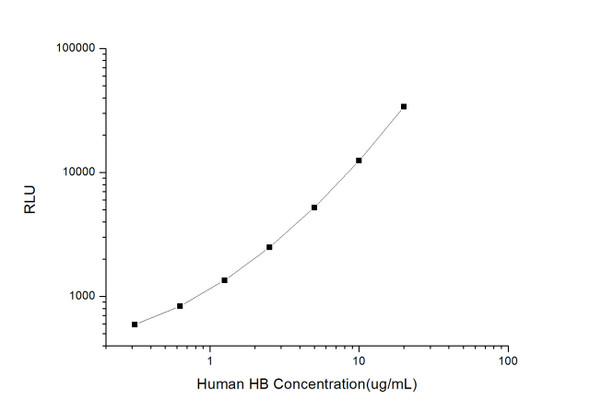
As the RLU values of the standard curve may vary according to the conditions of the actual assay performance (e. g. operator, pipetting technique, washing technique or temperature effects), the operator should establish a standard curve for each test. Typical standard curve and data is provided below for reference only.
| Concentration (µg/mL) | RLU | Average | Corrected |
| 20 | 31813 36151 | 33982 | 33950 |
| 10 | 12240 12700 | 12470 | 12438 |
| 5 | 5291 5209 | 5250 | 5218 |
| 2.5 | 2411 2637 | 2524 | 2492 |
| 1.25 | 1399 1367 | 1383 | 1351 |
| 0.63 | 895 839 | 867 | 835 |
| 0.31 | 617 629 | 623 | 591 |
| 0 | 32 32 | 32 | -- |
Precision
Intra-assay Precision (Precision within an assay): 3 samples with low, mid range and high level Human HB beta were tested 20 times on one plate, respectively.
Inter-assay Precision (Precision between assays): 3 samples with low, mid range and high level Human HB beta were tested on 3 different plates, 20 replicates in each plate.
| Intra-assay Precision | Inter-assay Precision | |||||
| Sample | 1 | 2 | 3 | 1 | 2 | 3 |
| n | 20 | 20 | 20 | 20 | 20 | 20 |
| Mean (µg/mL) | 0.98 | 3.03 | 8.01 | 1.03 | 2.84 | 7.54 |
| Standard deviation | 0.09 | 0.35 | 0.64 | 0.13 | 0.22 | 0.81 |
| C V (%) | 9.18 | 11.55 | 7.99 | 12.62 | 7.75 | 10.74 |
Recovery
The recovery of Human HB beta spiked at three different levels in samples throughout the range of the assay was evaluated in various matrices.
| Sample Type | Range (%) | Average Recovery (%) |
| Serum (n=5) | 86-99 | 91 |
| EDTA plasma (n=5) | 89-102 | 96 |
| Cell culture media (n=5) | 94-108 | 100 |
Linearity
Samples were spiked with high concentrations of Human HB beta and diluted with Reference Standard & Sample Diluent to produce samples with values within the range of the assay.
| Serum (n=5) | EDTA plasma (n=5) | Cell culture media (n=5) | ||
| 1:2 | Range (%) | 102-112 | 95-111 | 86-99 |
| Average (%) | 107 | 103 | 92 | |
| 1:4 | Range (%) | 91-105 | 85-98 | 93-109 |
| Average (%) | 98 | 92 | 99 | |
| 1:8 | Range (%) | 95-108 | 94-107 | 100-114 |
| Average (%) | 101 | 99 | 106 | |
| 1:16 | Range (%) | 89-102 | 102-118 | 95-111 |
| Average (%) | 96 | 110 | 102 |
An unopened kit can be stored at 4°C for 1 month. If the kit is not used within 1 month, store the items separately according to the following conditions once the kit is received.
| Item | Specifications | Storage |
| Micro CLIA Plate(Dismountable) | 8 wells ×12 strips | -20°C, 6 months |
| Reference Standard | 2 vials | |
| Concentrated Biotinylated Detection Ab (100×) | 1 vial, 120 µL | |
| Concentrated HRP Conjugate (100×) | 1 vial, 120 µL | -20°C(shading light), 6 months |
| Reference Standard & Sample Diluent | 1 vial, 20 mL | 4°C, 6 months |
| Biotinylated Detection Ab Diluent | 1 vial, 14 mL | |
| HRP Conjugate Diluent | 1 vial, 14 mL | |
| Concentrated Wash Buffer (25×) | 1 vial, 30 mL | |
| Substrate Reagent A | 1 vial, 5 mL | 4°C (shading light) |
| Substrate Reagent B | 1 vial, 5 mL | 4°C (shading light) |
| Plate Sealer | 5 pieces | |
| Product Description | 1 copy | |
| Certificate of Analysis | 1 copy |
- Set standard, test sample and control (zero) wells on the pre-coated plate and record theirpositions. It is recommended to measure each standard and sample in duplicate. Note: addall solutions to the bottom of the plate wells while avoiding contact with the well walls. Ensuresolutions do not foam when adding to the wells.
- Aliquot 100 µL of standard solutions into the standard wells.
- Add 100 µL of Sample / Standard dilution buffer into the control (zero) well.
- Add 100 µL of properly diluted sample (serum, plasma, tissue homogenates and otherbiological fluids. ) into test sample wells.
- Cover the plate with the sealer provided in the kit and incubate for 90 min at 37 °C.
- Aspirate the liquid from each well, do not wash. Immediately add 100 µL of BiotinylatedDetection Ab working solution to each well. Cover the plate with a plate seal and gently mix. Incubate for 1 hour at 37 °C.
- Aspirate or decant the solution from the plate and add 350 µL of wash buffer to each welland incubate for 1-2 minutes at room temperature. Aspirate the solution from each well andclap the plate on absorbent filter paper to dry. Repeat this process 3 times. Note: a microplatewasher can be used in this step and other wash steps.
- Add 100 µL of HRP Conjugate working solution to each well. Cover with a plate seal andincubate for 30 min at 37 °C.
- Aspirate or decant the solution from each well. Repeat the wash process for five times asconducted in step 7.
- Add 100 µL of Substrate mixture solution to each well. Cover with a new plate seal andincubate for no more than 5 min at 37 °C. Protect the plate from light.
- Determine the RLU value of each well immediately.

