Human Metabolism ELISA Kits
Human CYP1B1 (Cytochrome P450, family 1, subfamily B, polypeptide 1) CLIA Kit (HUES00275)
- SKU:
- HUES00275
- Product Type:
- ELISA Kit
- ELISA Type:
- CLIA Kit
- Size:
- 96 Assays
- Sensitivity:
- 18.75pg/mL
- Range:
- 31.25-2000pg/mL
- ELISA Type:
- Sandwich
- Synonyms:
- CP1B, CYPIB1, GLC3A, P4501B1
- Reactivity:
- Human
- Sample Type:
- Serum, plasma and other biological fluids
- Research Area:
- Metabolism
Description
| Assay type: | Sandwich |
| Format: | 96T |
| Assay time: | 4.5h |
| Reactivity: | Human |
| Detection method: | Chemiluminescence |
| Detection range: | 31.25-2000 pg/mL |
| Sensitivity: | 18.75 pg/mL |
| Sample volume: | 100µL |
| Sample type: | Tissue homogenates,cell lysates and other biological fluids |
| Repeatability: | CV < 15% |
| Specificity: | This kit recognizes Human CYP1B1 in samples. No significant cross-reactivity or interference between Human CYP1B1 and analogues was observed. |
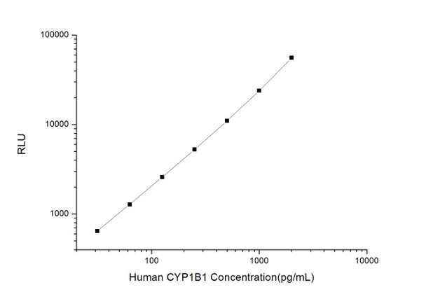
This kit uses Sandwich-CLIA as the method. The micro CLIA plate provided in this kit has been pre-coated with an antibody specific to Human CYP1B1. Standards or samples are added to the appropriate micro CLIA plate wells and combined with the specific antibody. Then a biotinylated detection antibody specific for Human CYP1B1 and Avidin-Horseradish Peroxidase (HRP) conjugate are added to each micro plate well successively and incubated. Free components are washed away. The substrate solution is added to each well. Only those wells that contain Human CYP1B1, biotinylated detection antibody and Avidin-HRP conjugate will appear fluorescence. The Relative light unit (RLU) value is measured spectrophotometrically by the Chemiluminescence immunoassay analyzer. The RLU value is positively associated with the concentration of Human CYP1B1. The concentration of Human CYP1B1 in the samples can be calculated by comparing the RLU of the samples to the standard curve.
| UniProt Protein Function: | CYP1B1: Cytochromes P450 are a group of heme-thiolate monooxygenases. In liver microsomes, this enzyme is involved in an NADPH-dependent electron transport pathway. It oxidizes a variety of structurally unrelated compounds, including steroids, fatty acids, and xenobiotics. Defects in CYP1B1 are the cause of primary congenital glaucoma type 3A (GLC3A). GLC3A is an autosomal recessive form of primary congenital glaucoma (PCG). PCG is characterized by marked increase of intraocular pressure at birth or early childhood, large ocular globes (buphthalmos) and corneal edema. It results from developmental defects of the trabecular meshwork and anterior chamber angle of the eye that prevent adequate drainage of aqueous humor. Defects in CYP1B1 are a cause of primary open angle glaucoma (POAG). POAG is a complex and genetically heterogeneous ocular disorder characterized by a specific pattern of optic nerve and visual field defects. The angle of the anterior chamber of the eye is open, and usually the intraocular pressure is increased. The disease is asymptomatic until the late stages, by which time significant and irreversible optic nerve damage has already taken place. In some cases, POAG shows digenic inheritance involving mutations in CYP1B1 and MYOC genes. Defects in CYP1B1 are a cause of Peters anomaly (PAN). Peters anomaly is a congenital defect of the anterior chamber of the eye. Belongs to the cytochrome P450 family. |
| UniProt Protein Details: | Protein type:Oxidoreductase; Xenobiotic Metabolism - metabolism by cytochrome P450; EC 1. 14. 14. 1; Amino Acid Metabolism - tryptophan Chromosomal Location of Human Ortholog: 2p22. 2 Cellular Component: endoplasmic reticulum membrane; mitochondrion Molecular Function:iron ion binding; heme binding; oxidoreductase activity, acting on paired donors, with incorporation or reduction of molecular oxygen, reduced flavin or flavoprotein as one donor, and incorporation of one atom of oxygen; oxygen binding; monooxygenase activity Biological Process: steroid metabolic process; estrogen metabolic process; collagen fibril organization; retinal metabolic process; positive regulation of apoptosis; response to toxin; positive regulation of JAK-STAT cascade; negative regulation of cell proliferation; visual perception; retinol metabolic process; arachidonic acid metabolic process; angiogenesis; nitric oxide biosynthetic process; cell adhesion; negative regulation of cell adhesion mediated by integrin; negative regulation of cell migration; epoxygenase P450 pathway; positive regulation of angiogenesis; inhibition of NF-kappaB transcription factor; xenobiotic metabolic process; toxin metabolic process; endothelial cell migration; blood vessel morphogenesis; aromatic compound metabolic process; membrane lipid catabolic process; induction of apoptosis by oxidative stress; sterol metabolic process Disease: Peters Anomaly; Glaucoma 3, Primary Congenital, A; Glaucoma 3, Primary Infantile, B |
| NCBI Summary: | This gene encodes a member of the cytochrome P450 superfamily of enzymes. The cytochrome P450 proteins are monooxygenases which catalyze many reactions involved in drug metabolism and synthesis of cholesterol, steroids and other lipids. The enzyme encoded by this gene localizes to the endoplasmic reticulum and metabolizes procarcinogens such as polycyclic aromatic hydrocarbons and 17beta-estradiol. Mutations in this gene have been associated with primary congenital glaucoma; therefore it is thought that the enzyme also metabolizes a signaling molecule involved in eye development, possibly a steroid. [provided by RefSeq, Jul 2008] |
| UniProt Code: | Q16678 |
| NCBI GenInfo Identifier: | 48429256 |
| NCBI Gene ID: | 1545 |
| NCBI Accession: | Q16678. 2 |
| UniProt Secondary Accession: | Q16678,Q5TZW8, Q93089, Q9H316, |
| UniProt Related Accession: | Q16678 |
| Molecular Weight: | |
| NCBI Full Name: | Cytochrome P450 1B1 |
| NCBI Synonym Full Names: | cytochrome P450, family 1, subfamily B, polypeptide 1 |
| NCBI Official Symbol: | CYP1B1 |
| NCBI Official Synonym Symbols: | CP1B; GLC3A; CYPIB1; P4501B1 |
| NCBI Protein Information: | cytochrome P450 1B1; microsomal monooxygenase; xenobiotic monooxygenase; aryl hydrocarbon hydroxylase; flavoprotein-linked monooxygenase; cytochrome P450, subfamily I (dioxin-inducible), polypeptide 1 (glaucoma 3, primary infantile) |
| UniProt Protein Name: | Cytochrome P450 1B1 |
| UniProt Synonym Protein Names: | CYPIB1 |
| Protein Family: | Cytochrome |
| UniProt Gene Name: | CYP1B1 |
| UniProt Entry Name: | CP1B1_HUMAN |
As the RLU values of the standard curve may vary according to the conditions of the actual assay performance (e. g. operator, pipetting technique, washing technique or temperature effects), the operator should establish a standard curve for each test. Typical standard curve and data is provided below for reference only.
| Concentration (pg/mL) | RLU | Average | Corrected |
| 2000 | 50226 61216 | 55721 | 55693 |
| 1000 | 22328 25620 | 23974 | 23946 |
| 500 | 11056 11008 | 11032 | 11004 |
| 250 | 4974 5614 | 5294 | 5266 |
| 125 | 2723 2493 | 2608 | 2580 |
| 62.5 | 1383 1239 | 1311 | 1283 |
| 31.25 | 659 689 | 674 | 646 |
| 0 | 27 29 | 28 | -- |
Precision
Intra-assay Precision (Precision within an assay): 3 samples with low, mid range and high level Human CYP1B1 were tested 20 times on one plate, respectively.
Inter-assay Precision (Precision between assays): 3 samples with low, mid range and high level Human CYP1B1 were tested on 3 different plates, 20 replicates in each plate.
| Intra-assay Precision | Inter-assay Precision | |||||
| Sample | 1 | 2 | 3 | 1 | 2 | 3 |
| n | 20 | 20 | 20 | 20 | 20 | 20 |
| Mean (pg/mL) | 106.19 | 291.22 | 905.74 | 115.25 | 310.56 | 945.08 |
| Standard deviation | 9.33 | 28.48 | 57.33 | 13.05 | 36.71 | 91.86 |
| C V (%) | 8.79 | 9.78 | 6.33 | 11.32 | 11.82 | 9.72 |
Recovery
The recovery of Human CYP1B1 spiked at three different levels in samples throughout the range of the assay was evaluated in various matrices.
| Sample Type | Range (%) | Average Recovery (%) |
| Serum (n=5) | 90-103 | 95 |
| EDTA plasma (n=5) | 84-99 | 90 |
| Cell culture media (n=5) | 85-100 | 92 |
Linearity
Samples were spiked with high concentrations of Human CYP1B1 and diluted with Reference Standard & Sample Diluent to produce samples with values within the range of the assay.
| Serum (n=5) | EDTA plasma (n=5) | Cell culture media (n=5) | ||
| 1:2 | Range (%) | 95-110 | 94-109 | 102-120 |
| Average (%) | 102 | 101 | 110 | |
| 1:4 | Range (%) | 94-106 | 104-117 | 85-99 |
| Average (%) | 101 | 110 | 92 | |
| 1:8 | Range (%) | 101-113 | 94-107 | 101-114 |
| Average (%) | 107 | 101 | 106 | |
| 1:16 | Range (%) | 92-107 | 92-109 | 100-115 |
| Average (%) | 99 | 99 | 107 |
An unopened kit can be stored at 4°C for 1 month. If the kit is not used within 1 month, store the items separately according to the following conditions once the kit is received.
| Item | Specifications | Storage |
| Micro CLIA Plate(Dismountable) | 8 wells ×12 strips | -20°C, 6 months |
| Reference Standard | 2 vials | |
| Concentrated Biotinylated Detection Ab (100×) | 1 vial, 120 µL | |
| Concentrated HRP Conjugate (100×) | 1 vial, 120 µL | -20°C(shading light), 6 months |
| Reference Standard & Sample Diluent | 1 vial, 20 mL | 4°C, 6 months |
| Biotinylated Detection Ab Diluent | 1 vial, 14 mL | |
| HRP Conjugate Diluent | 1 vial, 14 mL | |
| Concentrated Wash Buffer (25×) | 1 vial, 30 mL | |
| Substrate Reagent A | 1 vial, 5 mL | 4°C (shading light) |
| Substrate Reagent B | 1 vial, 5 mL | 4°C (shading light) |
| Plate Sealer | 5 pieces | |
| Product Description | 1 copy | |
| Certificate of Analysis | 1 copy |
- Set standard, test sample and control (zero) wells on the pre-coated plate and record theirpositions. It is recommended to measure each standard and sample in duplicate. Note: addall solutions to the bottom of the plate wells while avoiding contact with the well walls. Ensuresolutions do not foam when adding to the wells.
- Aliquot 100µl of standard solutions into the standard wells.
- Add 100µl of Sample / Standard dilution buffer into the control (zero) well.
- Add 100µl of properly diluted sample (serum, plasma, tissue homogenates and otherbiological fluids. ) into test sample wells.
- Cover the plate with the sealer provided in the kit and incubate for 90 min at 37°C.
- Aspirate the liquid from each well, do not wash. Immediately add 100µL of BiotinylatedDetection Ab working solution to each well. Cover the plate with a plate seal and gently mix. Incubate for 1 hour at 37°C.
- Aspirate or decant the solution from the plate and add 350µL of wash buffer to each welland incubate for 1-2 minutes at room temperature. Aspirate the solution from each well andclap the plate on absorbent filter paper to dry. Repeat this process 3 times. Note: a microplatewasher can be used in this step and other wash steps.
- Add 100µL of HRP Conjugate working solution to each well. Cover with a plate seal andincubate for 30 min at 37°C.
- Aspirate or decant the solution from each well. Repeat the wash process for five times asconducted in step 7.
- Add 100µL of Substrate mixture solution to each well. Cover with a new plate seal andincubate for no more than 5 min at 37°C. Protect the plate from light.
- Determine the RLU value of each well immediately.


