Human Cell Biology ELISA Kits 3
Human CD59 (Protectin) CLIA Kit (HUES00444)
- SKU:
- HUES00444
- Product Type:
- ELISA Kit
- ELISA Type:
- CLIA Kit
- Size:
- 96 Assays
- Sensitivity:
- 9.38pg/mL
- Range:
- 15.63-1000pg/mL
- ELISA Type:
- Sandwich
- Reactivity:
- Human
- Sample Type:
- Serum, plasma and other biological fluids
- Research Area:
- Cell Biology
Description
| Assay type: | Sandwich |
| Format: | 96T |
| Assay time: | 4.5h |
| Reactivity: | Human |
| Detection method: | Chemiluminescence |
| Detection range: | 15.63-1000 pg/mL |
| Sensitivity: | 9.38 pg/mL |
| Sample volume: | 100µL |
| Sample type: | Serum, plasma and other biological fluids |
| Repeatability: | CV < 15% |
| Specificity: | This kit recognizes Human CD59 in samples. No significant cross-reactivity or interference between Human CD59 and analogues was observed. |
This kit uses Sandwich-CLIA as the method. The micro CLIA plate provided in this kit has been pre-coated with an antibody specific to Human CD59. Standards or samples are added to the appropriate micro CLIA plate wells and combined with the specific antibody. Then a biotinylated detection antibody specific for Human CD59 and Avidin-Horseradish Peroxidase (HRP) conjugate are added to each micro plate well successively and incubated. Free components are washed away. The substrate solution is added to each well. Only those wells that contain Human CD59, biotinylated detection antibody and Avidin-HRP conjugate will appear fluorescence. The Relative light unit (RLU) value is measured spectrophotometrically by the Chemiluminescence immunoassay analyzer. The RLU value is positively associated with the concentration of Human CD59. The concentration of Human CD59 in the samples can be calculated by comparing the RLU of the samples to the standard curve.
| UniProt Protein Function: | CD59: Potent inhibitor of the complement membrane attack complex (MAC) action. Acts by binding to the C8 and/or C9 complements of the assembling MAC, thereby preventing incorporation of the multiple copies of C9 required for complete formation of the osmolytic pore. This inhibitor appears to be species-specific. Involved in signal transduction for T-cell activation complexed to a protein tyrosine kinase. Defects in CD59 are the cause of CD59 deficiency (CD59D). |
| UniProt Protein Details: | Protein type:Membrane protein, GPI anchor Chromosomal Location of Human Ortholog: 11p13 Cellular Component: anchored to external side of plasma membrane; cell surface; endoplasmic reticulum membrane; ER-Golgi intermediate compartment membrane; extracellular space; focal adhesion; Golgi membrane; membrane; plasma membrane; vesicle Molecular Function:complement binding; protein binding Biological Process: blood coagulation; cell activation; cell surface receptor linked signal transduction; COPII coating of Golgi vesicle; ER to Golgi vesicle-mediated transport; neutrophil degranulation; regulation of complement activation Disease: Hemolytic Anemia, Cd59-mediated, With Or Without Immune-mediated Polyneuropathy |
| NCBI Summary: | This gene encodes a cell surface glycoprotein that regulates complement-mediated cell lysis, and it is involved in lymphocyte signal transduction. This protein is a potent inhibitor of the complement membrane attack complex, whereby it binds complement C8 and/or C9 during the assembly of this complex, thereby inhibiting the incorporation of multiple copies of C9 into the complex, which is necessary for osmolytic pore formation. This protein also plays a role in signal transduction pathways in the activation of T cells. Mutations in this gene cause CD59 deficiency, a disease resulting in hemolytic anemia and thrombosis, and which causes cerebral infarction. Multiple alternatively spliced transcript variants, which encode the same protein, have been identified for this gene. [provided by RefSeq, Jul 2008] |
| UniProt Code: | P13987 |
| NCBI GenInfo Identifier: | 116021 |
| NCBI Gene ID: | 966 |
| NCBI Accession: | P13987. 1 |
| UniProt Related Accession: | P13987 |
| Molecular Weight: | 14kDa |
| NCBI Full Name: | CD59 glycoprotein |
| NCBI Synonym Full Names: | CD59 molecule (CD59 blood group) |
| NCBI Official Symbol: | CD59 |
| NCBI Official Synonym Symbols: | 1F5; EJ16; EJ30; EL32; G344; MIN1; MIN2; MIN3; MIRL; HRF20; MACIF; MEM43; MIC11; MSK21; 16. 3A5; HRF-20; MAC-IP; p18-20 |
| NCBI Protein Information: | CD59 glycoprotein |
| UniProt Protein Name: | CD59 glycoprotein |
| UniProt Synonym Protein Names: | 1F5 antigen; 20 kDa homologous restriction factor; HRF-20; HRF20; MAC-inhibitory protein; MAC-IP; MEM43 antigen; Membrane attack complex inhibition factor; MACIF; Membrane inhibitor of reactive lysis; MIRL; Protectin; CD_antigen: CD59 |
| Protein Family: | CD59 glycoprotein |
| UniProt Gene Name: | CD59 |
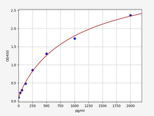
As the RLU values of the standard curve may vary according to the conditions of the actual assay performance (e. g. operator, pipetting technique, washing technique or temperature effects), the operator should establish a standard curve for each test. Typical standard curve and data is provided below for reference only.
| Concentration (pg/mL) | RLU | Average | Corrected |
| 1000 | 53113 54237 | 53675 | 53650 |
| 500 | 22228 22750 | 22489 | 22464 |
| 250 | 11000 9422 | 10211 | 10186 |
| 125 | 4743 5059 | 4901 | 4876 |
| 62.5 | 2466 2440 | 2453 | 2428 |
| 31.25 | 1299 1263 | 1281 | 1256 |
| 15.63 | 682 734 | 708 | 683 |
| 0 | 24 26 | 25 | -- |
Precision
Intra-assay Precision (Precision within an assay): 3 samples with low, mid range and high level Human CD59 were tested 20 times on one plate, respectively.
Inter-assay Precision (Precision between assays): 3 samples with low, mid range and high level Human CD59 were tested on 3 different plates, 20 replicates in each plate.
| Intra-assay Precision | Inter-assay Precision | |||||
| Sample | 1 | 2 | 3 | 1 | 2 | 3 |
| n | 20 | 20 | 20 | 20 | 20 | 20 |
| Mean (pg/mL) | 54.60 | 120.88 | 475.96 | 54.10 | 109.30 | 441.40 |
| Standard deviation | 5.40 | 13.20 | 50.02 | 5.46 | 10.83 | 48.29 |
| C V (%) | 9.89 | 10.92 | 10.51 | 10.09 | 9.91 | 10.94 |
Recovery
The recovery of Human CD59 spiked at three different levels in samples throughout the range of the assay was evaluated in various matrices.
| Sample Type | Range (%) | Average Recovery (%) |
| Serum (n=5) | 100-117 | 108 |
| EDTA plasma (n=5) | 96-111 | 104 |
| Cell culture media (n=5) | 100-117 | 107 |
Linearity
Samples were spiked with high concentrations of Human CD59 and diluted with Reference Standard & Sample Diluent to produce samples with values within the range of the assay.
| Serum (n=5) | EDTA plasma (n=5) | Cell culture media (n=5) | ||
| 1:2 | Range (%) | 88-103 | 92-110 | 89-103 |
| Average (%) | 95 | 100 | 95 | |
| 1:4 | Range (%) | 89-101 | 89-101 | 94-107 |
| Average (%) | 96 | 96 | 99 | |
| 1:8 | Range (%) | 93-107 | 87-99 | 95-107 |
| Average (%) | 99 | 93 | 102 | |
| 1:16 | Range (%) | 91-108 | 83-95 | 84-99 |
| Average (%) | 99 | 90 | 90 |
An unopened kit can be stored at 4°C for 1 month. If the kit is not used within 1 month, store the items separately according to the following conditions once the kit is received.
| Item | Specifications | Storage |
| Micro CLIA Plate(Dismountable) | 8 wells ×12 strips | -20°C, 6 months |
| Reference Standard | 2 vials | |
| Concentrated Biotinylated Detection Ab (100×) | 1 vial, 120 µL | |
| Concentrated HRP Conjugate (100×) | 1 vial, 120 µL | -20°C(shading light), 6 months |
| Reference Standard & Sample Diluent | 1 vial, 20 mL | 4°C, 6 months |
| Biotinylated Detection Ab Diluent | 1 vial, 14 mL | |
| HRP Conjugate Diluent | 1 vial, 14 mL | |
| Concentrated Wash Buffer (25×) | 1 vial, 30 mL | |
| Substrate Reagent A | 1 vial, 5 mL | 4°C (shading light) |
| Substrate Reagent B | 1 vial, 5 mL | 4°C (shading light) |
| Plate Sealer | 5 pieces | |
| Product Description | 1 copy | |
| Certificate of Analysis | 1 copy |
- Set standard, test sample and control (zero) wells on the pre-coated plate and record theirpositions. It is recommended to measure each standard and sample in duplicate. Note: addall solutions to the bottom of the plate wells while avoiding contact with the well walls. Ensuresolutions do not foam when adding to the wells.
- Aliquot 100µl of standard solutions into the standard wells.
- Add 100µl of Sample / Standard dilution buffer into the control (zero) well.
- Add 100µl of properly diluted sample (serum, plasma, tissue homogenates and otherbiological fluids. ) into test sample wells.
- Cover the plate with the sealer provided in the kit and incubate for 90 min at 37°C.
- Aspirate the liquid from each well, do not wash. Immediately add 100µL of BiotinylatedDetection Ab working solution to each well. Cover the plate with a plate seal and gently mix. Incubate for 1 hour at 37°C.
- Aspirate or decant the solution from the plate and add 350µL of wash buffer to each welland incubate for 1-2 minutes at room temperature. Aspirate the solution from each well andclap the plate on absorbent filter paper to dry. Repeat this process 3 times. Note: a microplatewasher can be used in this step and other wash steps.
- Add 100µL of HRP Conjugate working solution to each well. Cover with a plate seal andincubate for 30 min at 37°C.
- Aspirate or decant the solution from each well. Repeat the wash process for five times asconducted in step 7.
- Add 100µL of Substrate mixture solution to each well. Cover with a new plate seal andincubate for no more than 5 min at 37°C. Protect the plate from light.
- Determine the RLU value of each well immediately.


