Description
GenieHTS Calcium Flux Assay Kit
High-throughput, no wash calcium (Ca²⁺) assay. An optimal solution for measuring intracellular Ca²⁺ dynamics, including effectors such as GPCRs and protein transporters.
| Product Name: | GenieHTS Calcium Flux Assay Kit |
| Product Code: | ASIB0001 |
| Product Size: | 10 plates |
| Excitation: | 490 nm |
| Emission: | 515 nm |
| Molecular Weight: | 1097 |
| Component Name | Size | Storage |
| GenieHTS Calcium Reagent | Lyophilized (10) | -20°C |
| DMSO | 225μL | 4°C |
| Dye Solvent | 4mL | 4°C |
| 10X Assay Buffer | 20mL | 4°C |
| TRS | 4mL | 4°C |
| Probenecid Solution | 4mL | 4°C |
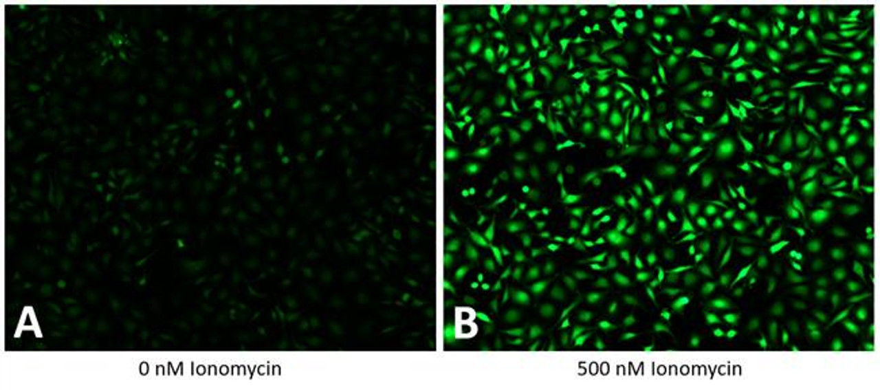
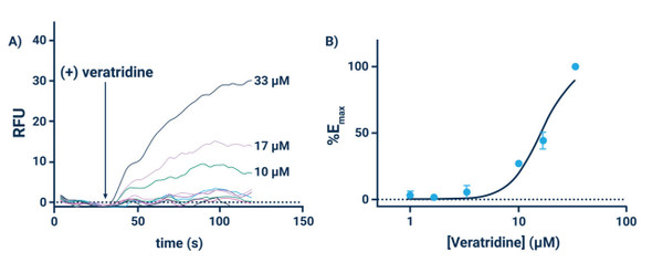
The GenieHTS Calcium Flux Assay Kit is a total assay solution for multi-well plate-based, high-throughput measurements of changes intracellular Ca2+ mediated through a wide-variety of plasma membrane and intracellular calcium channels and transporters. The Assay Genie GenieHTS Calcium Flux Assay is also useful for investigating numerous effectors of ion channels and transporters including G protein-coupled receptors, lipid kinases and protein kinases. In multi- well, plate-based formats, the GenieHTS Calcium Flux Assay can be used to discover and characterize the effects of many tens-of-thousands of compounds and environmental factors on effectors of intracellular Ca2+. For the last 25 years, fluorescence-based measures of Ca2+ flux have brought about the discovery of small-molecule modulators of a host of Ion channels, transporters, GPCRS and other targets of interest for both drug discovery and basic research.
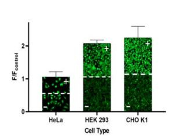
The Assay Genie GenieHTS Calcium Flux Assay provides all the reagents necessary for use as a washed or no-wash assay with adherent or non-adherent cells. The optional use of a probenecid solution and an extracellular background masking solution offers the ultimate in compatibility for cells types which are difficult to load with fluorescent Ca2+ indicators (e.g. Chinese Hamster Ovary, CHO cells) and when performing assays in complete, serum-containing cell culture medium is desired.
Wash Method Adherent Cells
The instructions given below are for one, 384-well microplate. Certain aspects of the instructions may need to be altered, as appropriate, for multiple microplates or 2other assay formats (e.g. 96-well microplates or non- adherent cells). The Assay Genie GenieHTS Calcium Flux indicator and Calcium indicator-containing solutions should be protected from direct light.
- Add 20 μL DMSO to the tube containing GenieHTS Calcium Reagent.
- Vortex until the GenieHTS Calcium Reagent is fully dissolved.
- Add water according to the below table to a 15 mL centrifuge tube.
- Add 1 mL of 10X Assay Buffer to tube from step 3.
- Add 200 μL of Dye Solvent to the tube from step 4.
- If desired add 200 μL of Probenecid Solution to the tube from step 5.
- Add 20 μL of GenieHTS Calcium Indicator Solution from step 2 to the tube from step 6.
- Briefly vortex the tube from step 7 to mix.
- Remove the cell-culture medium from the 384-well microplate containing the cells of interest.
- Add 20 μL per well of the Dye Loading Solution from step 8 to the microplate from step 9.
- Incubate the microplate containing the cells and Dye Loading Solution for 1 hour at 37°C.
- Briefly vortex the tube from step 12 to mix.
- Remove Dye Loading Solution from microplate in step 11.
- Add 20 μL per well of the Wash Solution prepared in step 13 to the microplate from step 14.
- Transfer the washed, dye-loaded, cell-containing microplate from step 15, along with an additional microplate containing a stimulus solution of interest, to a kinetic-imaging plate reader (e.g. WaveFront Panoptic, Hamamatsu FDSS, Molecular Devices FLIPR or Molecular Devices FlexStation).
- Acquire data using an excitation wavelength of ~ 490 nm, an emission wavelength of ~ 520 nm and an acquisition frequency of 1 Hz. Begin data acquisition and after 10 seconds add 5 μL of the stimulus solution to the cell-containing plate and continue data acquisition for an additional 90 seconds.**
**The timing of and volume of stimulus solution addition may vary. Some experiments may include the addition of other solutions to the cell-containing microplate prior to the addition of the stimulus solution. In these cases, the volume of the stimulus solution addition should be altered to account for the additional volume of solution in the cell-containing microplate.
Dye Loading Solution (Wash Method)
| Component | Method 1 | Method 2 |
| GenieHTS Calcium Indicator Solution | 20μL | 20μL |
| Dye Solvent | 200μL | 200μL |
| 10X Assay Buffer | 1mL | 1mL |
| Probenecid Solution* | - | 200μL |
| Water | 8.8mL | 8.6mL |
| Total | 10mL | 10mL |
* Probenecid may be included in the Dye Loading Solution to aid dye retention. This may be particularly important in certain cell lines (e.g. CHO cells). However, caution is advised when using Probenecid as it may have undesirable effects on assay performance for the target of interest.
Wash Solution (Wash Method)
| Component | Method 1 | Method 2 | Method 3 | Method 4 |
| 10X Assay Buffer | 1mL | 1mL | 1mL | 1mL |
| TRS* | - | 200μL | - | 200μL |
| Probenecid Solution | - | - | 200μL | 200μL |
| Water | 9mL | 8.8mL | 8.8mL | 8.6mL |
| Total | 10mL | 10mL | 10mL | 10mL |
*TRS contains a membrane-impermeant dye useful for masking extracellular fluorescence. Caution is advised when using TRS or other extracellular masking solutions as they may have undesirable effects on assay performance for the target of interest.
No-wash Method — Adherent Cells
- Add 20 μL DMSO to the tube containing GenieHTS Calcium Indicator.
- Vortex until the GenieHTS Calcium Indicator is fully dissolved.
- Add appropriate volume of water (Table 4) to a 15 mL centrifuge tube.
- Add 1 mL of 10X Assay Buffer to tube from step 3.
- Add 400 μL of Dye Solvent to the tube from step 4.
- Add 400 μL of TRS to the tube from step 5.
- If desired add 400 μL of Probenecid Solution to the tube from step 6.
- Add 20 μL of GenieHTS Calcium Indicator Solution from step 2 to the tube from step 7.
- Briefly vortex the tube from step 8 to mix.
- Add 20 μL per well of the Dye Loading Solution from step 9 to the cell-containing microplate. Do not remove the cell culture medium.
- Incubate the microplate containing the cells and Dye Loading Solution for 1 hour at 37°C in a cell culture incubator.
- Transfer the dye-loaded, cell-containing microplate from step 11, along with an additional microplate containing a stimulus solution of interest, to a kinetic-imaging plate reader (e.g. WaveFront Panoptic, Hamamatsu FDSS, Molecular Devices FLIPR or Molecular Devices FlexStation).
- Acquire data using an excitation wavelength of ~ 490 nm, an emission wavelength of ~ 520 nm and an acquisition frequency of 1 Hz. Begin data acquisition and after 10 seconds add 10 μL of the stimulus solution to the cell-containing plate and continue data acquisition for an additional 90 seconds.**
**The timing of and volume of stimulus solution addition may vary. Some experiments may include the addition of other solutions to the cell-containing microplate prior to the addition of the stimulus solution. In these cases, the volume of the stimulus solution addition should be altered to account for the additional volume of solution in the cell-containing microplate.
Dye Loading Solution (No-wash Method)
| Component | Method 1 | Method 2 |
| GenieHTS Calcium Indicator Solution | 20μL | 20μL |
| Dye Solvent | 400μL | 400μL |
| 10X Assay Buffer | 1mL | 1mL |
| TRS* | 400μL | 400μL |
| Probenecid Solution*** | - | 400μL |
| Water | 8.2mL | 7.8mL |
| Total | 10mL | 10mL |
*TRS contains a membrane-impermeant dye useful for masking extracellular fluorescence. Caution is advised when using TRS or other extracellular masking solutions as they may have undesirable effects on assay performance for the target of interest.
**Probenecid may be included in the Dye Loading Solution to aid dye retention. This may be particularly important in certain cell lines (e.g. CHO cells). However, caution is advised when using Probenecid as it may have undesirable effects on assay.


