Developmental Biology
Anti-YY1 Antibody (CAB19569)
- SKU:
- CAB19569
- Product Type:
- Antibody
- Reactivity:
- Human
- Reactivity:
- Mouse
- Reactivity:
- Rat
- Host Species:
- Rabbit
- Isotype:
- IgG
- Research Area:
- Developmental Biology
Description
| Antibody Name: | Anti-YY1 Antibody |
| Antibody SKU: | CAB19569 |
| Antibody Size: | 20uL, 50uL, 100uL |
| Application: | WB IF |
| Reactivity: | Human, Mouse, Rat |
| Host Species: | Rabbit |
| Immunogen: | A synthesized peptide derived from human YY1. |
| Application: | WB IF |
| Recommended Dilution: | WB 1:500 - 1:2000 IF 1:50 - 1:200 |
| Reactivity: | Human, Mouse, Rat |
| Positive Samples: | Mouse liver, Mouse kidney, Mouse spleen, Rat spleen, U-251MG, HeLa, Jurkat, Raji |
| Immunogen: | A synthesized peptide derived from human YY1. |
| Purification Method: | Affinity purification |
| Storage Buffer: | Store at -20°C. Avoid freeze / thaw cycles. Buffer: PBS with 0.02% sodium azide, 0.05% BSA, 50% glycerol, pH7.3. |
| Isotype: | IgG |
| Sequence: | Email for sequence |
| Gene ID: | 7528 |
| Uniprot: | P25490 |
| Cellular Location: | |
| Calculated MW: | 68kDa |
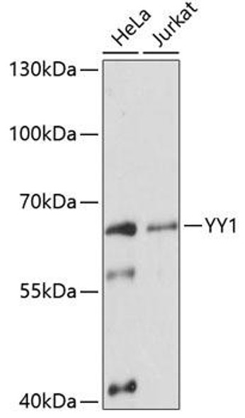
| Observed MW: | 68KDa |
 | Western blot analysis of extracts of various cell lines, using YY1 antibody at 1:1000 dilution. Secondary antibody: HRP Goat Anti-Rabbit IgG (H+L) at 1:10000 dilution. Lysates/proteins: 25ug per lane. Blocking buffer: 3% nonfat dry milk in TBST. Detection: ECL Basic Kit. Exposure time: 10s. |
 | Western blot analysis of extracts of various cell lines, using YY1 antibody at 1:1000 dilution. Secondary antibody: HRP Goat Anti-Rabbit IgG (H+L) at 1:10000 dilution. Lysates/proteins: 25ug per lane. Blocking buffer: 3% nonfat dry milk in TBST. Detection: ECL Basic Kit. Exposure time: 1s. |
 | Immunofluorescence analysis of C6 cells using YY1 antibody (A19569). Blue: DAPI for nuclear staining. |
 | Immunoprecipitation analysis of 300ug extracts of HeLa cells using 3ug YY1 antibody at a dilition of 1:2000. |
 | Chromatin immunoprecipitation analysis of extracts of 293T cells, using YY1 antibody and rabbit IgG. The amount of immunoprecipitated DNA was checked by quantitative PCR. Histogram was constructed by the ratios of the immunoprecipitated DNA to the input. |


