Description
| Antibody Name: | Anti-YKL-40/CHI3L1 Antibody (CAB20792) |
| Antibody SKU: | CAB20792 |
| Antibody Size: | 50µL, 100µL |
| Application: | Western blotting, Immunofluorescence |
| Reactivity: | Human, Mouse |
| Host Species: | Rabbit |
| Immunogen: | A synthetic peptide corresponding to a sequence within amino acids 284-383 of human YKL-40/CHI3L1 (NP_001267.2). |
| Application: | Western blotting, Immunofluorescence |
| Recommended Dilution: | WB 1:500 - 1:2000 IF 1:50 - 1:200 |
| Reactivity: | Human, Mouse |
| Positive Samples: | THP-1, U-87MG, Mouse lung |
| Immunogen: | A synthetic peptide corresponding to a sequence within amino acids 284-383 of human YKL-40/CHI3L1 (NP_001267.2). |
| Purification Method: | Affinity purification |
| Storage Buffer: | Store at -20°C. Avoid freeze / thaw cycles. Buffer: PBS with 0.02% sodium azide, 50% glycerol, pH7.3. |
| Isotype: | IgG |
| Sequence: | PGRF TKEA GTLA YYEI CDFL RGAT VHRI LGQQ VPYA TKGN QWVG YDDQ ESVK SKVQ YLKD RQLA GAMV WALD LDDF QGSF CGQD LRFP LTNA IKDA LAAT |
| Cellular Location: | Cytoplasm, Endoplasmic reticulum, Secreted, extracellular space, perinuclear region |
| Calculated MW: | 42kDa |
| Observed MW: | 30-43KDa |
| Synonyms: | CHI3L1, ASRT7, CGP-39, GP-39, GP39, HC-gp39, HCGP-3P, YKL-40, YKL40, YYL-40, hCGP-39 |
| Background: | Chitinases catalyze the hydrolysis of chitin, which is an abundant glycopolymer found in insect exoskeletons and fungal cell walls. The glycoside hydrolase 18 family of chitinases includes eight human family members. This gene encodes a glycoprotein member of the glycosyl hydrolase 18 family. The protein lacks chitinase activity and is secreted by activated macrophages, chondrocytes, neutrophils and synovial cells. The protein is thought to play a role in the process of inflammation and tissue remodeling. |
 | Immunofluorescence analysis of THP-1 and K562 cells using YKL-40/CHI3L1 Rabbit mAb at dilution of 1:100 (40x lens). Blue: DAPI for nuclear staining. |
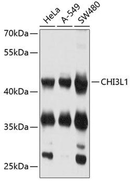
 | Western blot analysis of extracts of various cell lines, using YKL-40/CHI3L1 antibody at 1:1000 dilution. Secondary antibody: HRP Goat Anti-Rabbit IgG (H+L) at 1:10000 dilution. Lysates/proteins: 25ug per lane. Blocking buffer: 3% nonfat dry milk in TBST. Detection: ECL Basic Kit. Exposure time: 90s. |
 | Immunofluorescence analysis of THP-1 and K562 cells using YKL-40/CHI3L1 Rabbit mAb at dilution of 1:100 (40x lens). Blue: DAPI for nuclear staining. |
 | Western blot analysis of extracts of THP-1 cells, using YKL-40/CHI3L1 antibody at 1:1000 dilution. Secondary antibody: HRP Goat Anti-Rabbit IgG (H+L) at 1:10000 dilution. Lysates/proteins: 25ug per lane. Blocking buffer: 3% nonfat dry milk in TBST. Detection: ECL Basic Kit. Exposure time: 1s. |


