Cell Biology Antibodies 14
Anti-VCAM1 Antibody (CAB16999)
- SKU:
- CAB16999
- Product Type:
- Antibody
- Reactivity:
- Human
- Reactivity:
- Mouse
- Reactivity:
- Rat
- Host Species:
- Rabbit
- Isotype:
- IgG
Description
| Antibody Name: | Anti-VCAM1 Antibody (CAB16999) |
| Antibody SKU: | CAB16999 |
| Antibody Size: | 50µL, 100µL |
| Application: | Western blotting, Immunofluorescence, Immunoprecipitation |
| Reactivity: | Human, Mouse, Rat |
| Host Species: | Rabbit |
| Immunogen: | Recombinant fusion protein containing a sequence corresponding to amino acids 480-630 of human VCAM1 (NP_001069.1). |
| Application: | Western blotting, Immunofluorescence, Immunoprecipitation |
| Recommended Dilution: | WB 1:500 - 1:2000 IF 1:50 - 1:200 IP 1:50 - 1:200 |
| Reactivity: | Human, Mouse, Rat |
| Positive Samples: | K-562, RAW264.7, Rat lung |
| Immunogen: | Recombinant fusion protein containing a sequence corresponding to amino acids 480-630 of human VCAM1 (NP_001069.1). |
| Purification Method: | Affinity purification |
| Storage Buffer: | Store at -20°C. Avoid freeze / thaw cycles. Buffer: PBS with 0.02% sodium azide, 50% glycerol, pH7.3. |
| Isotype: | IgG |
| Sequence: | ALVC QAKL HIDD MEFE PKQR QSTQ TLYV NVAP RDTT VLVS PSSI LEEG SSVN MTCL SQGF PAPK ILWS RQLP NGEL QPLS ENAT LTLI STKM EDSG VYLC EGIN QAGR SRKE VELI IQVT PKDI KLTA FPSE SVKE GDTV IISC TCGN VPE |
| Cellular Location: | Membrane, Single-pass type I membrane protein |
| Calculated MW: | 71kDa/74kDa/81kDa |
| Observed MW: | 85-100KDa |
| Synonyms: | CD106, INCAM-100, VCAM1 |
| Background: | This gene is a member of the Ig superfamily and encodes a cell surface sialoglycoprotein expressed by cytokine-activated endothelium. This type I membrane protein mediates leukocyte-endothelial cell adhesion and signal transduction, and may play a role in the development of artherosclerosis and rheumatoid arthritis. Three alternatively spliced transcripts encoding different isoforms have been described for this gene. |
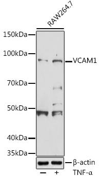
 | Western blot analysis of extracts of RAW264. 7 cells, using VCAM1 antibody at 1:500 dilution. RAW264. 7 cells were treated by TNF-α (20 ng/ml) at 37℃ for 16 hours after serum-starvation overnight. Secondary antibody: HRP Goat Anti-Rabbit IgG (H+L) at 1:10000 dilution. Lysates/proteins: 25ug per lane. Blocking buffer: 3% nonfat dry milk in TBST. Detection: ECL Basic Kit. Exposure time: 180s. |
 | Western blot analysis of extracts of various cell lines, using VCAM1 antibody at 1:500 dilution. Secondary antibody: HRP Goat Anti-Rabbit IgG (H+L) at 1:10000 dilution. Lysates/proteins: 25ug per lane. Blocking buffer: 3% nonfat dry milk in TBST. Detection: ECL Basic Kit. Exposure time: 90s. |


