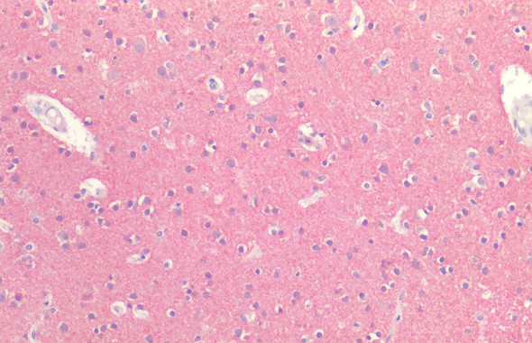
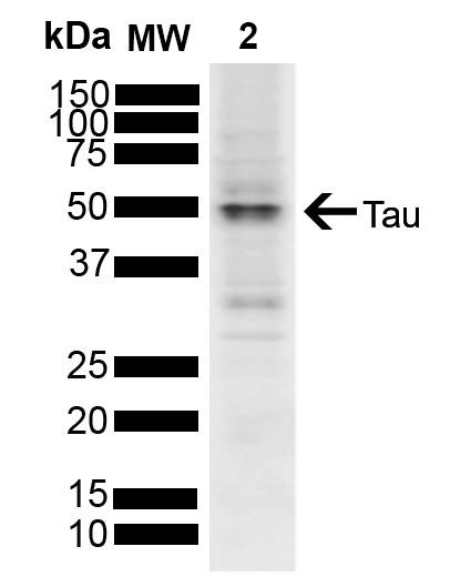
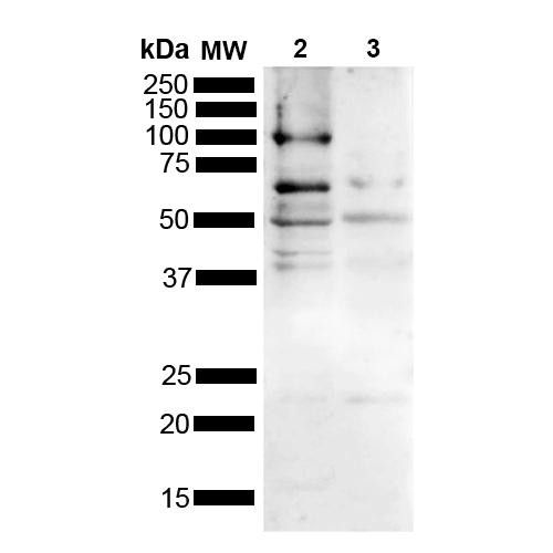
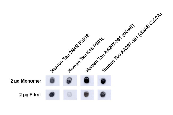
Tau Antibody [1D5] is a mouse monoclonal antibody directed against Tau, offered by Assay Genie for research applications. Reported applications include WB, DB, ICC/IF and ELISA. Reported reactivity: Human, Mouse and Rat. Supplied as IgG1, purified by Protein G.
Tau is a microtubule-associated protein predominantly found in neuronal axons, where it stabilizes microtubules and supports axonal transport. In the adult human brain, six isoforms of tau are expressed, generated by alternative splicing of the MAPT gene. These isoforms contain either three (3R) or four (4R) microtubule-binding repeat domains, with 2N4R (Tau-441) representing the full-length form. In healthy neurons, tau is tightly regulated. However, in neurodegenerative diseases known as tauopathies—including Alzheimer’s disease (AD), frontotemporal dementia, and progressive supranuclear palsy—tau becomes abnormally hyperphosphorylated. This modification reduces its affinity for microtubules, leading to the formation of insoluble neurofibrillary tangles (NFTs), a pathological hallmark of AD. Mutations in the MAPT gene, such as P301S (encoded by exon 10), impair tau’s ability to bind microtubules and promote aggregation. These mutations are commonly used in transgenic models to study tau-mediated neurotoxicity, synaptic dysfunction, and neuronal death. Tau pathology correlates strongly with cognitive decline in AD and is increasingly recognized as a driver of disease progression. As such, tau is a major focus of biomarker development and therapeutic targeting in neurodegenerative research. This antibody is also available conjugated to ATTO 390, ATTO 488, ATTO 594, APC, Biotin, FITC, HRP, PerCP and RPE. Supplied in 100 µg. For research use only; not for diagnostic or therapeutic procedures.
Tau, TAU, TAU_HUMAN, MAPT, MAPTL, Microtubule-associated protein tau, Microtubule associated protein tau, Microtubule associated protein tau isoform 4, Neurofibrillary tangle protein, Paired helical filament tau, Paired helical filament-tau, PHF tau, PHF-tau, DDPAC, FTDP 17, G protein beta1/gamma2 subunit interacting factor 1, MSTD, Mtapt, MTBT1, MTBT2, PPND, PPP1R103, Protein phosphatase 1 regulatory subunit 103, RNPTAU, AI413597, AW045860, FLJ31424, MGC134287, MGC138549, MGC156663
References:
1. www.alz.org/alzheimers-dementia/facts-figures 2. Alzheimer, A. Über eine eigenartige Erkrankung der Hirnrinde. Allg. Z. Psychiatr. Psych.-Gerichtl. Med. 64, 146–148 (1907) 3. Matsumoto, G. et al. (2018). Int J Mol Sci. 19, 1497. 4. Goedert, M. and Spillantini, M. G. (2017). Mol Brain. 10:18. 5. Bugiani, O. et al. (1999). J Neuropathol Exp Neurol. 58(6):667-77.


![Anti-EpCAM (1D5) [1D5-B6-B10] Monoclonal Antibody (AGMB04359)](https://cdn11.bigcommerce.com/s-rd6ounxcu2/images/stencil/590x590/products/116308/130346/anti-epcam-1d5-1d5-b6-b10-monoclonal-antibody-agmb04359__78313__17791.1786847270.jpg?c=1 "Anti-EpCAM (1D5) [1D5-B6-B10] Monoclonal Antibody (AGMB04359)")

![Anti-DDB2 [R09-1D5] Monoclonal Antibody (AGMB00491)](https://cdn11.bigcommerce.com/s-rd6ounxcu2/images/stencil/590x590/products/112441/126489/anti-ddb2-r09-1d5-monoclonal-antibody-agmb00491__05075__97991.1786837348.jpg?c=1 "Anti-DDB2 [R09-1D5] Monoclonal Antibody (AGMB00491)")