Anti-TAF15 Antibody (CAB9383)
")
- SKU:
- CAB9383
- Product Type:
- Antibody
- Antibody Type:
- Monoclonal Antibody
- Reactivity:
- Human
- Reactivity:
- Mouse
- Reactivity:
- Rat
- Host Species:
- Rabbit
- Isotype:
- IgG
")
Description
| Antibody Name: | Anti-TAF15 Antibody (CAB9383) |
| Antibody SKU: | CAB9383 |
| Antibody Size: | 50µL, 100µL |
| Application: | Western blotting, Immunohistochemistry, Immunofluorescence, Immunoprecipitation |
| Reactivity: | Human, Mouse, Rat |
| Host Species: | Rabbit |
| Immunogen: | A synthesized peptide derived from human TAF15. |
| Application: | Western blotting, Immunohistochemistry, Immunofluorescence, Immunoprecipitation |
| Recommended Dilution: | WB 1:500 - 1:2000 IHC 1:50 - 1:200 IF 1:50 - 1:200 IP 1:50 - 1:200 |
| Reactivity: | Human, Mouse, Rat |
| Positive Samples: | HeLa, 293T, HT-29, Mouse thymus |
| Immunogen: | A synthesized peptide derived from human TAF15. |
| Purification Method: | Affinity purification |
| Storage Buffer: | Store at -20°C. Avoid freeze / thaw cycles. Buffer: PBS with 0.02% sodium azide, 50% glycerol, pH7.3. |
| Isotype: | IgG |
| Sequence: | Email for sequence |
| Cellular Location: | Cytoplasm, Nucleus |
| Calculated MW: | 75kDa |
| Observed MW: | 75KDa |
| Synonyms: | TAF15, Npl3, RBP56, TAF2N, TAFII68, TATA-binding protein-associated factor 2N |
| Background: | This gene encodes a member of the TET family of RNA-binding proteins. The encoded protein plays a role in RNA polymerase II gene transcription as a component of a distinct subset of multi-subunit transcription initiation factor TFIID complexes. Translocations involving this gene play a role in acute leukemia and extraskeletal myxoid chondrosarcoma, and mutations in this gene may play a role in amyotrophic lateral sclerosis. Alternatively spliced transcript variants encoding multiple isoforms have been observed for this gene. |
 | Immunofluorescence analysis of NIH/3T3 cells using TAF15 antibody at dilution of 1:25. Blue: DAPI for nuclear staining. |
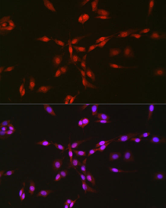 | Immunofluorescence analysis of PC-12 cells using TAF15 antibody at dilution of 1:25. Blue: DAPI for nuclear staining. |
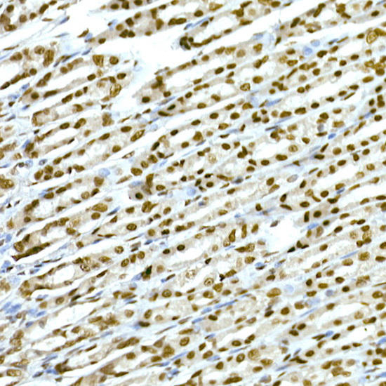 | Immunohistochemistry of paraffin-embedded rat stomach using TAF15 Rabbit mAb at dilution of 1:25 (40x lens). Perform high pressure antigen retrieval with 10 mM citrate buffer pH 6. 0 before commencing with IHC staining protocol. |
 | Immunofluorescence analysis of HeLa cells using TAF15 antibody at dilution of 1:25. Blue: DAPI for nuclear staining. |
 | Immunohistochemistry of paraffin-embedded rat lung using TAF15 Rabbit mAb at dilution of 1:25 (40x lens). Perform high pressure antigen retrieval with 10 mM citrate buffer pH 6. 0 before commencing with IHC staining protocol. |
 | Immunohistochemistry of paraffin-embedded mouse testis using TAF15 Rabbit mAb at dilution of 1:25 (40x lens). Perform high pressure antigen retrieval with 10 mM citrate buffer pH 6. 0 before commencing with IHC staining protocol. |
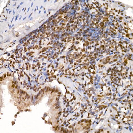
 | Immunohistochemistry of paraffin-embedded human tonsil using TAF15 Rabbit mAb at dilution of 1:25 (40x lens). Perform high pressure antigen retrieval with 10 mM citrate buffer pH 6. 0 before commencing with IHC staining protocol. |
 | Immunohistochemistry of paraffin-embedded human lung cancer using TAF15 Rabbit mAb at dilution of 1:25 (40x lens). Perform high pressure antigen retrieval with 10 mM citrate buffer pH 6. 0 before commencing with IHC staining protocol. |
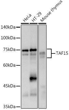 | Western blot analysis of extracts of various cell lines, using at 1:500 dilution. Secondary antibody: HRP Goat Anti-Rabbit IgG (H+L) at 1:10000 dilution. Lysates/proteins: 25ug per lane. Blocking buffer: 3% nonfat dry milk in TBST. Detection: ECL Basic Kit. Exposure time: 1s. |
Related Products
")
")

")
 ELISA Kit")
Human anti-EPO antibody(anti-Erythropoietin antibody) ELISA Kit
system_update_altDatasheet - तकनीकी मैनुअलsystem_update_altMSDS - तकनीक