Anti-Symmetric DiMethyl-Histone H3-R8 Antibody (CAB20820)
(No reviews yet)
Write a Review
")
- SKU:
- CAB20820
")
Description
| Antibody Name: | Anti-Symmetric DiMethyl-Histone H3-R8 Antibody (CAB20820) |
| Antibody SKU: | CAB20820 |
| Antibody Size: | 50µL, 100µL |
| Application: | Western blotting |
| Reactivity: | Human, Mouse, Rat |
| Host Species: | Rabbit |
| Immunogen: | A synthetic symmetric dimethylated peptide around R8 of human histone H3 (NP_003520.1). |
| Application: | Western blotting |
| Recommended Dilution: | WB 1:500 - 1:2000 ChIP 1:50 - 1:200 |
| Reactivity: | Human, Mouse, Rat |
| Positive Samples: | HeLa, NIH/3T3, C6, H3 |
| Immunogen: | A synthetic symmetric dimethylated peptide around R8 of human histone H3 (NP_003520.1). |
| Purification Method: | Affinity purification |
| Storage Buffer: | Store at -20°C. Avoid freeze / thaw cycles. Buffer: PBS with 0.02% sodium azide, 50% glycerol, pH7.3. |
| Isotype: | IgG |
| Sequence: | QTAR K |
| Cellular Location: | Chromosome, Nucleus |
| Calculated MW: | 15kDa |
| Observed MW: | 17KDa |
| Synonyms: | H3/A, H3C2, H3C3, H3C4, H3C6, H3C7, H3C8, H3FA, H3C10, H3C11, H3C12, HIST1H3A |
| Background: | Histones are basic nuclear proteins that are responsible for the nucleosome structure of the chromosomal fiber in eukaryotes. This structure consists of approximately 146 bp of DNA wrapped around a nucleosome, an octamer composed of pairs of each of the four core histones (H2A, H2B, H3, and H4). The chromatin fiber is further compacted through the interaction of a linker histone, H1, with the DNA between the nucleosomes to form higher order chromatin structures. This gene is intronless and encodes a replication-dependent histone that is a member of the histone H3 family. Transcripts from this gene lack polyA tails; instead, they contain a palindromic termination element. This gene is found in the large histone gene cluster on chromosome 6p22-p21.3. |
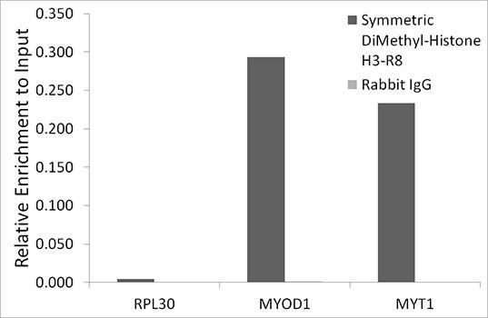 | Chromatin immunoprecipitation analysis of extracts of HeLa cells, using Symmetric DiMethyl-Histone H3-R8 antibody and rabbit IgG. The amount of immunoprecipitated DNA was checked by quantitative PCR. Histogram was constructed by the ratios of the immunoprecipitated DNA to the input. |
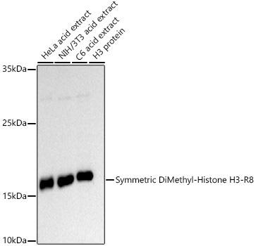 | Western blot analysis of extracts of various cell lines, using Symmetric DiMethyl-Histone H3-R8 antibody at 1:500 dilution. Secondary antibody: HRP Goat Anti-Rabbit IgG (H+L) at 1:10000 dilution. Lysates/proteins: 25ug per lane. Blocking buffer: 3% nonfat dry milk in TBST. Detection: ECL Basic Kit. Exposure time: 90s. |
Additional Information
Product Type: |
Antibody |
Antibody Type: |
Secondary Antibody |
Host Species: |
Rabbit |
Isotype: |
IgG |
Related Products
")
Anti-Symmetric DiMethyl-Histone H3-R8 Antibody (CAB2374)
Epigenetics & Nuclear Signaling Antibodies 3
")
Symmetric DiMethyl-Histone H3-R8 Monoclonal Antibody (CAB21207)
OverviewProduct Name:Symmetric DiMethyl-Histone H3-R8 Monoclonal AntibodyProduct Code:CAB21207Reactivity:Human, Mouse, Rat, Other (Wide Range)
")
Symmetric DiMethyl-Histone H3-R8 Monoclonal Antibody (CAB22363)
Symmetric DiMethyl-Histone H3-R8 Monoclonal Antibody (CAB22363)The product page for this antibody is being prepared. If you have a question please don't hesitate to get in touch!
")
Anti-Asymmetric DiMethyl-Histone H3-R8 Antibody (CAB3157)
Epigenetics & Nuclear Signaling Antibodies 3
")
Anti-Symmetric DiMethyl-Histone H3-R17 Antibody (CAB3152)
Epigenetics & Nuclear Signaling Antibodies 3