Description
| Antibody Name: | Anti-Prdm9 Antibody (CAB20832) |
| Antibody SKU: | CAB20832 |
| Antibody Size: | 50µL, 100µL |
| Application: | Western blotting, Immunohistochemistry |
| Reactivity: | Human, Mouse, Rat |
| Host Species: | Rabbit |
| Immunogen: | Recombinant fusion protein containing a sequence corresponding to amino acids 87-370 of mouse Prdm9 (NP_659058.3). |
| Application: | Western blotting, Immunohistochemistry |
| Recommended Dilution: | WB 1:500 - 1:2000 IHC 1:50 - 1:200 |
| Reactivity: | Human, Mouse, Rat |
| Positive Samples: | A-549, Mouse testis, Rat testis, Rat kidney |
| Immunogen: | Recombinant fusion protein containing a sequence corresponding to amino acids 87-370 of mouse Prdm9 (NP_659058.3). |
| Purification Method: | Affinity purification |
| Storage Buffer: | Store at -20°C. Avoid freeze / thaw cycles. Buffer: PBS with 0.02% sodium azide, 50% glycerol, pH7.3. |
| Isotype: | IgG |
| Sequence: | DSED SDEE WTPK QQVS PPWV PFRV KHSK QQKE SSRM PFSG ESNV KEGS GIEN LLNT SGSE HVQK PVSS LEEG NTSG QHSG KKLK LRKK NVEV KMYR LRER KGLA YEEV SEPQ DDDY LYCE KCQN FFID SCPN HGPP LFVK DSMV DRGH PNHS VLSL PPGL RISP SGIP EAGL GVWN EASD LPVG LHFG PYEG QITE DEEA ANSG YSWL ITKG RNCY EYVD GQDE SQAN WMRY VNCA RDDE EQNL VAFQ YHRK IFYR TCRV IRPG CELL VWYG DEYG QELG IKWG |
| Cellular Location: | |
| Calculated MW: | 97kDa |
| Observed MW: | 110KDa |
| Synonyms: | Rc, re, Dsbc, Rcr1, Dsbc1, Meise, repro7, Meisetz, PRDM9-B, BC012016, G1-419-29 |
| Background: |
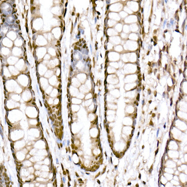
 | Immunohistochemistry of paraffin-embedded human colon using Prdm9 Rabbit mAb at dilution of 1:800 (40x lens). Perform high pressure antigen retrieval with 10 mM citrate buffer pH 6. 0 before commencing with IHC staining protocol. |
 | Western blot analysis of extracts of Rat kidney, using Prdm9 antibody at 1:1000 dilution. Secondary antibody: HRP Goat Anti-Rabbit IgG (H+L) at 1:10000 dilution. Lysates/proteins: 25ug per lane. Blocking buffer: 3% nonfat dry milk in TBST. Detection: ECL Basic Kit. Exposure time: 30s. |
 | Immunohistochemistry of paraffin-embedded human colon carcinoma using Prdm9 Rabbit mAb at dilution of 1:800 (40x lens). Perform high pressure antigen retrieval with 10 mM citrate buffer pH 6. 0 before commencing with IHC staining protocol. |
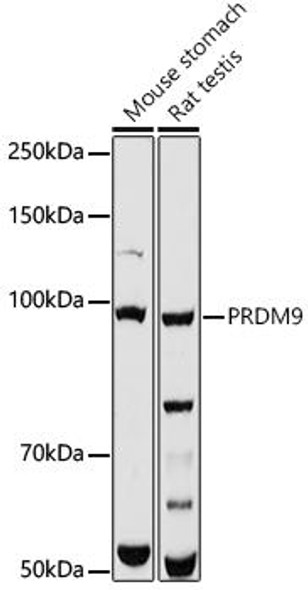
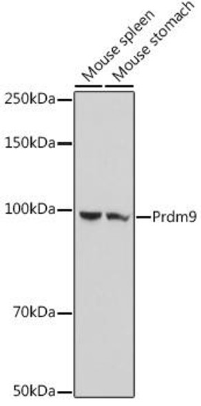
 | Western blot analysis of extracts of various cell lines, using Prdm9 antibody at 1:1000 dilution. Secondary antibody: HRP Goat Anti-Rabbit IgG (H+L) at 1:10000 dilution. Lysates/proteins: 25ug per lane. Blocking buffer: 3% nonfat dry milk in TBST. Detection: ECL Basic Kit. Exposure time: 10s. |


