Description
| Antibody Name: | Anti-PLK1 Antibody (CAB21082) |
| Antibody SKU: | CAB21082 |
| Antibody Size: | 50µL, 100µL |
| Application: | Western blotting, Immunohistochemistry |
| Reactivity: | Human, Mouse, Rat |
| Host Species: | Rabbit |
| Immunogen: | Recombinant fusion protein containing a sequence corresponding to amino acids 272-495 of human PLK1 (NP_005021.2). |
| Application: | Western blotting, Immunohistochemistry |
| Recommended Dilution: | WB 1:500 - 1:2000 IHC 1:50 - 1:200 |
| Reactivity: | Human, Mouse, Rat |
| Positive Samples: | HeLa, Jurkat, K-562, COS-7, Mouse testis, Rat testis |
| Immunogen: | Recombinant fusion protein containing a sequence corresponding to amino acids 272-495 of human PLK1 (NP_005021.2). |
| Purification Method: | Affinity purification |
| Storage Buffer: | Store at -20°C. Avoid freeze / thaw cycles. Buffer: PBS with 0.02% sodium azide, 50% glycerol, pH7.3. |
| Isotype: | IgG |
| Sequence: | KHIN PVAA SLIQ KMLQ TDPT ARPT INEL LNDE FFTS GYIP ARLP ITCL TIPP RFSI APSS LDPS NRKP LTVL NKGL ENPL PERP REKE EPVV RETG EVVD CHLS DMLQ QLHS VNAS KPSE RGLV RQEE AEDP ACIP IFWV SKWV DYSD KYGL GYQL CDNS VGVL FNDS TRLI LYND GDSL QYIE RDGT ESYL TVSS HPNS LMKK ITLL KYFR NYMS EHLL KAGA |
| Cellular Location: | Chromosome, Cytoplasm, Midbody, Nucleus, centromere, centrosome, cytoskeleton, kinetochore, microtubule organizing center, spindle |
| Calculated MW: | 68kDa |
| Observed MW: | 62kDa |
| Synonyms: | PLK1, PLK, STPK13 |
| Background: | The Ser/Thr protein kinase encoded by this gene belongs to the CDC5/Polo subfamily. It is highly expressed during mitosis and elevated levels are found in many different types of cancer. Depletion of this protein in cancer cells dramatically inhibited cell proliferation and induced apoptosis; hence, it is a target for cancer therapy. |
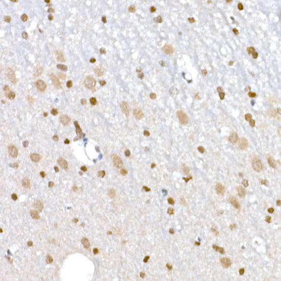 | Immunohistochemistry of paraffin-embedded rat spleen using PLK1 Rabbit mAb at dilution of 1:100 (40x lens). Perform high pressure antigen retrieval with 10 mM citrate buffer pH 6. 0 before commencing with IHC staining protocol. |
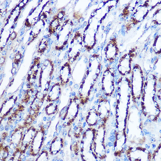 | Immunohistochemistry of paraffin-embedded mouse spleen using PLK1 Rabbit mAb at dilution of 1:100 (40x lens). Perform high pressure antigen retrieval with 10 mM citrate buffer pH 6. 0 before commencing with IHC staining protocol. |
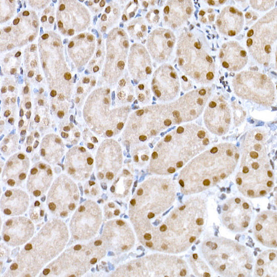 | Immunohistochemistry of paraffin-embedded rat ovary using PLK1 Rabbit mAb at dilution of 1:100 (40x lens). Perform high pressure antigen retrieval with 10 mM citrate buffer pH 6. 0 before commencing with IHC staining protocol. |
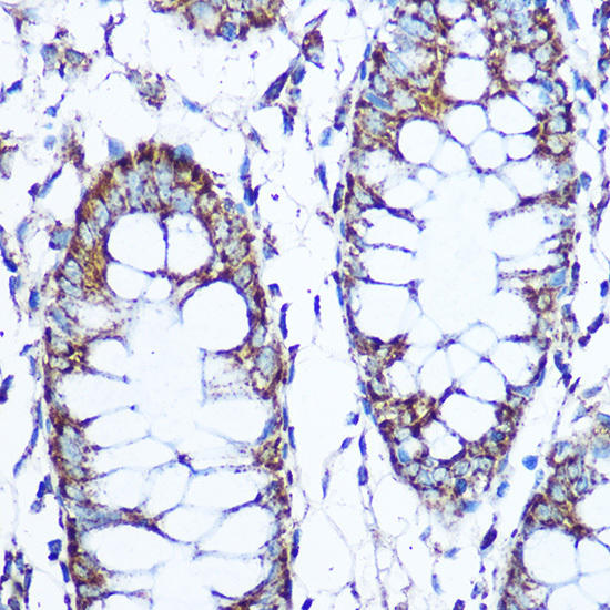 | Immunohistochemistry of paraffin-embedded human tonsil using PLK1 Rabbit mAb at dilution of 1:100 (40x lens). Perform high pressure antigen retrieval with 10 mM citrate buffer pH 6. 0 before commencing with IHC staining protocol. |
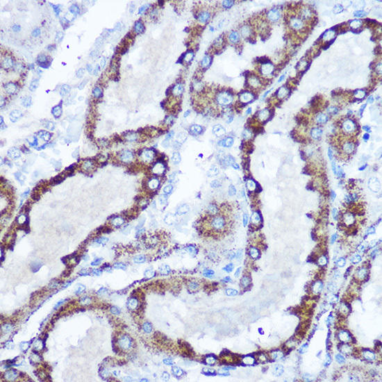 | Immunohistochemistry of paraffin-embedded human esophageal cancer using PLK1 Rabbit mAb at dilution of 1:100 (40x lens). Perform high pressure antigen retrieval with 10 mM citrate buffer pH 6. 0 before commencing with IHC staining protocol. |
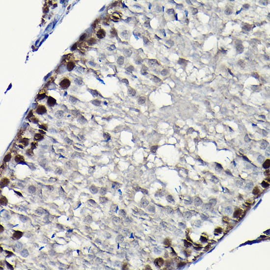 | Immunohistochemistry of paraffin-embedded human colon carcinoma using PLK1 Rabbit mAb at dilution of 1:100 (40x lens). Perform high pressure antigen retrieval with 10 mM citrate buffer pH 6. 0 before commencing with IHC staining protocol. |
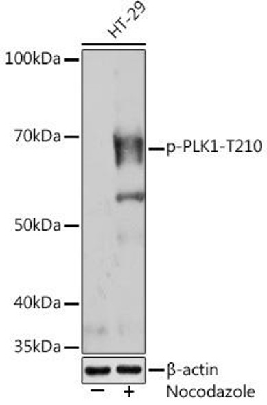
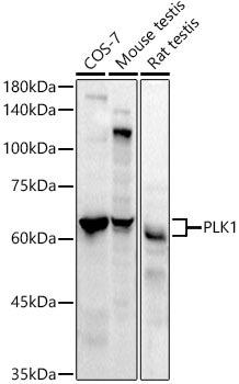 | Western blot analysis of extracts of various cell lines, using PLK1 antibody at 1:1000 dilution. Secondary antibody: HRP Goat Anti-Rabbit IgG (H+L) at 1:10000 dilution. Lysates/proteins: 25ug per lane. Blocking buffer: 3% nonfat dry milk in TBST. Detection: ECL Basic Kit. Exposure time: 60s. |
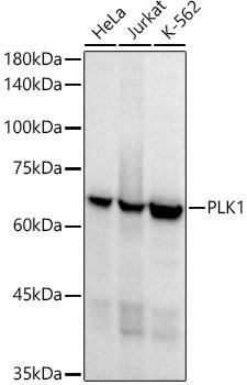 | Western blot analysis of extracts of various cell lines, using PLK1 antibody at 1:1000 dilution. Secondary antibody: HRP Goat Anti-Rabbit IgG (H+L) at 1:10000 dilution. Lysates/proteins: 25ug per lane. Blocking buffer: 3% nonfat dry milk in TBST. Detection: ECL Basic Kit. Exposure time: 10s. |


