Description
| Antibody Name: | Anti-PKM1-specific Antibody (CAB21052) |
| Antibody SKU: | CAB21052 |
| Antibody Size: | 50µL, 100µL |
| Application: | Western blotting |
| Reactivity: | Human, Mouse, Rat |
| Host Species: | Rabbit |
| Immunogen: | A synthetic peptide corresponding to a sequence within amino acids 350-450 of human PKM1 (NP_001193728.1). |
| Application: | Western blotting |
| Recommended Dilution: | WB 1:500 - 1:2000 |
| Reactivity: | Human, Mouse, Rat |
| Positive Samples: | RD, Mouse skeletal muscle, Rat skeletal muscle, Rat brain |
| Immunogen: | A synthetic peptide corresponding to a sequence within amino acids 350-450 of human PKM1 (NP_001193728.1). |
| Purification Method: | Affinity purification |
| Storage Buffer: | Store at -20°C. Avoid freeze / thaw cycles. Buffer: PBS with 0.02% sodium azide, 50% glycerol, pH7.3. |
| Isotype: | IgG |
| Sequence: | GSDV ANAV LDGA DCIM LSGE TAKG DYPL EAVR MQHL IARE AEAA MFHR KLFE ELVR ASSH STDL MEAM AMGS VEAS YKCL AAAL IVLT ESGR SAHQ VARY R |
| Cellular Location: | Cytoplasm, Nucleus |
| Calculated MW: | 58kDa |
| Observed MW: | 60KDa |
| Synonyms: | CTHBP, HEL-S-30, OIP3, PK3, PKM2, TCB, THBP1, PKM |
| Background: | This gene encodes a protein involved in glycolysis. The encoded protein is a pyruvate kinase that catalyzes the transfer of a phosphoryl group from phosphoenolpyruvate to ADP, generating ATP and pyruvate. This protein has been shown to interact with thyroid hormone and may mediate cellular metabolic effects induced by thyroid hormones. This protein has been found to bind Opa protein, a bacterial outer membrane protein involved in gonococcal adherence to and invasion of human cells, suggesting a role of this protein in bacterial pathogenesis. Several alternatively spliced transcript variants encoding a few distinct isoforms have been reported. provided by RefSeq, May 2011 |
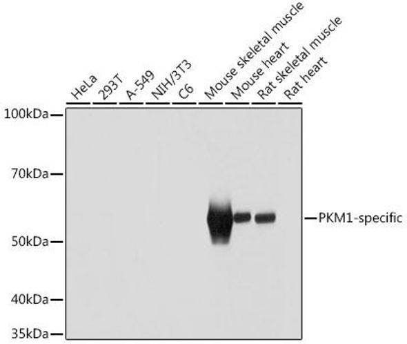
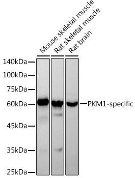 | Western blot analysis of extracts of various cell lines, using PKM1-specific antibody at 1:1000 dilution. Secondary antibody: HRP Goat Anti-Rabbit IgG (H+L) at 1:10000 dilution. Lysates/proteins: 25ug per lane. Blocking buffer: 3% nonfat dry milk in TBST. Detection: ECL Basic Kit. Exposure time: 1s. |
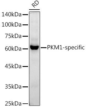 | Western blot analysis of extracts of RD cells, using PKM1-specific antibody at 1:1000 dilution. Secondary antibody: HRP Goat Anti-Rabbit IgG (H+L) at 1:10000 dilution. Lysates/proteins: 25ug per lane. Blocking buffer: 3% nonfat dry milk in TBST. Detection: ECL Basic Kit. Exposure time: 60s. |


