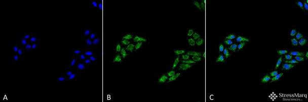
p53 Antibody is a rabbit polyclonal antibody directed against p53, offered by Assay Genie for research applications. Reported applications include WB and ICC/IF. Reported reactivity: Human. Purified by Peptide Affinity.
p53 (tumor protein 53) is a transcription factor best known for its role in tumor suppression, but it also plays a critical role in neuronal survival, DNA repair, and apoptosis. Often referred to as the “guardian of the genome,” p53 maintains genomic stability by regulating cell cycle checkpoints and initiating cell death in response to DNA damage. In the brain, p53 is activated by oxidative stress, excitotoxicity, and mitochondrial dysfunction—key features of neurodegenerative diseases. Overactivation of p53 can lead to excessive neuronal apoptosis, contributing to the progression of Alzheimer’s, Parkinson’s, and Huntington’s disease. Targeting p53 signaling pathways offers a promising strategy to modulate neuronal death and enhance neuroprotection in degenerative conditions. This antibody is also available conjugated to ATTO 390, ATTO 488, ATTO 594, APC, Biotin, FITC, HRP, PerCP and RPE. Supplied in 100 µg. For research use only; not for diagnostic or therapeutic procedures.
Product Name:
p53 Antibody
Product SKU:
STPA00069
Size:
100 µg
Target:
p53
Host Species:
Rabbit
Antibody Type:
Polyclonal
Research Areas:
Cancer, Cell Cycle, p53 Pathway, Cancer, Tumor suppressors, Apoptosis, Cell Signaling, Epigenetics and Nuclear Signaling, DNA/RNA, DNA Damage and Repair
Buffer:
PBS, 50% glycerol, 0.09% sodium azide
Storage & Shipping:
Store at -20°C. Shipped on Blue Ice or 4°C. Conjugated variants should be stored according to the product label.
Format:
Liquid
Applications:
WB, ICC/IF
Purification:
Peptide Affinity Purified
Concentration:
1 mg/ml
Reactivity:
Human
Recommended Dilution:
WB (1:1000); ICC/IF (1:100); optimal dilutions for assays should be determined by the user.
Specificity:
Predicted molecular weight at ~43.7kDa. Observed bands at ~53kDa.
Guarantee:
12 months from date of dispatch
Immunogen:
Synthetic peptide from the C-terminal of human tumor protein p53.
TP53, Tumor Protein 53, BCC7, LFS1, TRP53, p53 tumor suppressor
References:
1. Georescu C.V., Saftoiu A., Georgescu C.C., Ciurea R. and Ciurea T. (2007) J Gastrointestin Liver Dis. 16(2): 133-9. 2. Strachan T., et al. (1999) Human Molecular Genetics 2. Ch. 18: Cancer Genetics. 3. Hansen J.E., et al. (2007) Cancer Research 67: 1769-1774. 4. Di Cesare E., et al. (2001) Ann Clin Lab Sci 31: 253-8. 5. Akere A., Otegbayo J.A. (2007) Singapore Med J. 48(1): 41-4.


![Anti-p53 [R07-6A7] Monoclonal Antibody (AGMB01752)](https://cdn11.bigcommerce.com/s-rd6ounxcu2/images/stencil/590x590/products/113699/127740/anti-p53-r07-6a7-monoclonal-antibody-agmb01752__55210__01886.1786840565.jpg?c=1 "Anti-p53 [R07-6A7] Monoclonal Antibody (AGMB01752)")
![Anti-p53 [R09-5E6] Monoclonal Antibody (AGMB01753)](https://cdn11.bigcommerce.com/s-rd6ounxcu2/images/stencil/590x590/products/113704/127745/anti-p53-r09-5e6-monoclonal-antibody-agmb01753__64404__86371.1786840577.jpg?c=1 "Anti-p53 [R09-5E6] Monoclonal Antibody (AGMB01753)")
![Anti-p53 [R07-5G9] Monoclonal Antibody (AGMB01754)](https://cdn11.bigcommerce.com/s-rd6ounxcu2/images/stencil/590x590/products/113698/127747/anti-p53-r07-5g9-monoclonal-antibody-agmb01754__89340__63636.1786840580.jpg?c=1 "Anti-p53 [R07-5G9] Monoclonal Antibody (AGMB01754)")
![Anti-p53 [R01-2B9] Monoclonal Antibody (AGMB02275)](https://cdn11.bigcommerce.com/s-rd6ounxcu2/images/stencil/590x590/products/114224/128265/anti-p53-r01-2b9-monoclonal-antibody-agmb02275__36825__09594.1786841915.jpg?c=1 "Anti-p53 [R01-2B9] Monoclonal Antibody (AGMB02275)")
![Anti-p53 [7J8-X3-J6] Monoclonal Antibody (AGMB06191)](https://cdn11.bigcommerce.com/s-rd6ounxcu2/images/stencil/590x590/products/118130/132169/anti-p53-7j8-x3-j6-monoclonal-antibody-agmb06191__03203__97301.1786851937.jpg?c=1 "Anti-p53 [7J8-X3-J6] Monoclonal Antibody (AGMB06191)")