Description
| Antibody Name: | Anti-NAT1 Antibody (CAB20897) |
| Antibody SKU: | CAB20897 |
| Antibody Size: | 50µL, 100µL |
| Application: | Western blotting, Immunofluorescence |
| Reactivity: | Human, Mouse, Rat |
| Host Species: | Rabbit |
| Immunogen: | Recombinant protein of Human NAT1. |
| Application: | Western blotting, Immunofluorescence |
| Recommended Dilution: | WB 1:500 - 1:2000 IF 1:50 - 1:200 |
| Reactivity: | Human, Mouse, Rat |
| Positive Samples: | HeLa, Jurkat, SH-SY5Y |
| Immunogen: | Recombinant protein of Human NAT1. |
| Purification Method: | Affinity purification |
| Storage Buffer: | Store at -20°C. Avoid freeze / thaw cycles. Buffer: PBS with 0.02% sodium azide, 0.05% BSA, 50% glycerol, pH7.3. |
| Isotype: | IgG |
| Sequence: | Email for sequence |
| Cellular Location: | Cytoplasm |
| Calculated MW: | 33kDa |
| Observed MW: | 33KDa |
| Synonyms: | NAT1, AAC1, MNAT, NAT-1, NATI |
| Background: | This gene is one of two arylamine N-acetyltransferase (NAT) genes in the human genome, and is orthologous to the mouse and rat Nat2 genes. The enzyme encoded by this gene catalyzes the transfer of an acetyl group from acetyl-CoA to various arylamine and hydrazine substrates. This enzyme helps metabolize drugs and other xenobiotics, and functions in folate catabolism. Multiple transcript variants encoding different isoforms have been found for this gene. |
 | Immunofluorescence analysis of PC-3 cells using NAT1 Rabbit mAb at dilution of 1:200 (40x lens). Blue: DAPI for nuclear staining. |
 | Immunofluorescence analysis of PC-12 cells using NAT1 Rabbit mAb at dilution of 1:200 (40x lens). Blue: DAPI for nuclear staining. |
 | Immunofluorescence analysis of NIH/3T3 cells using NAT1 Rabbit mAb at dilution of 1:200 (40x lens). Blue: DAPI for nuclear staining. |
 | Immunofluorescence analysis of HepG2 cells using NAT1 Rabbit mAb at dilution of 1:200 (40x lens). Blue: DAPI for nuclear staining. |
 | Immunofluorescence analysis of HeLa cells using NAT1 Rabbit mAb at dilution of 1:200 (40x lens). Blue: DAPI for nuclear staining. |
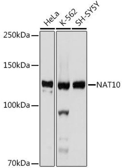
 | Western blot analysis of extracts of various cell lines, using NAT1 antibody at 1:1000 dilution. Secondary antibody: HRP Goat Anti-Rabbit IgG (H+L) at 1:10000 dilution. Lysates/proteins: 25ug per lane. Blocking buffer: 3% nonfat dry milk in TBST. Detection: ECL Enhanced Kit. Exposure time: 180s. |


