Description
| Antibody Name: | Anti-MYL12B Antibody (CAB9387) |
| Antibody SKU: | CAB9387 |
| Antibody Size: | 50µL, 100µL |
| Application: | Western blotting, Immunofluorescence |
| Reactivity: | Human, Mouse, Rat |
| Host Species: | Rabbit |
| Immunogen: | A synthesized peptide derived from human MYL12B. |
| Application: | Western blotting, Immunofluorescence |
| Recommended Dilution: | WB 1:500 - 1:2000 IF 1:50 - 1:200 |
| Reactivity: | Human, Mouse, Rat |
| Positive Samples: | K-562, U-87MG, Mouse large intestine, Mouse lung, Mouse kidney, Rat lung, Rat brain |
| Immunogen: | A synthesized peptide derived from human MYL12B. |
| Purification Method: | Affinity purification |
| Storage Buffer: | Store at -20°C. Avoid freeze / thaw cycles. Buffer: PBS with 0.02% sodium azide, 50% glycerol, pH7.3. |
| Isotype: | IgG |
| Sequence: | Email for sequence |
| Cellular Location: | |
| Calculated MW: | 18kDa |
| Observed MW: | 20KDa |
| Synonyms: | MYL12B, MLC-B, MRLC2, myosin light chain 12B |
| Background: | The activity of nonmuscle myosin II (see MYH9; MIM 160775) is regulated by phosphorylation of a regulatory light chain, such as MRLC2. This phosphorylation results in higher MgATPase activity and the assembly of myosin II filaments (Iwasaki et al., 2001 PubMed 11942626).supplied by OMIM, Mar 2008 |
 | Western blot analysis of extracts of various cell lines, using at 1:500 dilution. Secondary antibody: HRP Goat Anti-Rabbit IgG (H+L) at 1:10000 dilution. Lysates/proteins: 25ug per lane. Blocking buffer: 3% nonfat dry milk in TBST. Detection: ECL Basic Kit. Exposure time: 90s. |
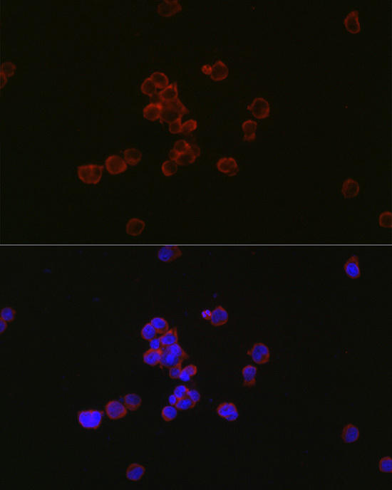
 | Immunofluorescence analysis of Jurkat cells using MYL12B antibody at dilution of 1:100. Blue: DAPI for nuclear staining. |
 | Western blot analysis of extracts of various cell lines, using at 1:500 dilution. Secondary antibody: HRP Goat Anti-Rabbit IgG (H+L) at 1:10000 dilution. Lysates/proteins: 25ug per lane. Blocking buffer: 3% nonfat dry milk in TBST. Detection: ECL Basic Kit. Exposure time: 3s. |


