Anti-MRPS31 Antibody (CAB20932)
")
- SKU:
- CAB20932
- Product Type:
- Antibody
- Antibody Type:
- Polyclonal Antibody
- Reactivity:
- Plant
- Host Species:
- Rabbit
- Isotype:
- IgG
")
Description
| Antibody Name: | Anti-MRPS31 Antibody (CAB20932) |
| Antibody SKU: | CAB20932 |
| Antibody Size: | 50µL, 100µL |
| Application: | Western blotting |
| Reactivity: | Human, Mouse, Rat |
| Host Species: | Rabbit |
| Immunogen: | Recombinant protein of Human MRPS31. |
| Application: | Western blotting |
| Recommended Dilution: | WB 1:500 - 1:2000 |
| Reactivity: | Human, Mouse, Rat |
| Positive Samples: | Jurkat, HepG2, HeLa, Mouse heart, Rat liver |
| Immunogen: | Recombinant protein of Human MRPS31. |
| Purification Method: | Affinity purification |
| Storage Buffer: | Store at -20°C. Avoid freeze / thaw cycles. Buffer: PBS with 0.02% sodium azide, 0.05% BSA, 50% glycerol, pH7.3. |
| Isotype: | IgG |
| Sequence: | Email for sequence |
| Cellular Location: | Mitochondrion |
| Calculated MW: | 45kDa |
| Observed MW: | 38KDa |
| Synonyms: | MRPS31, IMOGN38, MRP-S31, S31mt |
| Background: | Mammalian mitochondrial ribosomal proteins are encoded by nuclear genes and help in protein synthesis within the mitochondrion. Mitochondrial ribosomes (mitoribosomes) consist of a small 28S subunit and a large 39S subunit. They have an estimated 75% protein to rRNA composition compared to prokaryotic ribosomes, where this ratio is reversed. Another difference between mammalian mitoribosomes and prokaryotic ribosomes is that the latter contain a 5S rRNA. Among different species, the proteins comprising the mitoribosome differ greatly in sequence, and sometimes in biochemical properties, which prevents easy recognition by sequence homology. The 28S subunit of the mammalian mitoribosome may play a crucial and characteristic role in translation initiation. This gene encodes a 28S subunit protein that has also been associated with type 1 diabetes; however, its relationship to the etiology of this disease remains to be clarified. Pseudogenes corresponding to this gene have been found on chromosomes 3 and 13. |
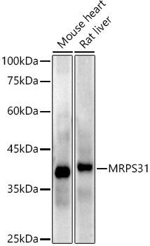 | Western blot analysis of extracts of various cell lines, using MRPS31 antibody at 1:1000 dilution. Secondary antibody: HRP Goat Anti-Rabbit IgG (H+L) at 1:10000 dilution. Lysates/proteins: 25ug per lane. Blocking buffer: 3% nonfat dry milk in TBST. Detection: ECL Enhanced Kit. Exposure time: 180s. |
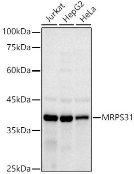 | Western blot analysis of extracts of various cell lines, using MRPS31 antibody at 1:1000 dilution. Secondary antibody: HRP Goat Anti-Rabbit IgG (H+L) at 1:10000 dilution. Lysates/proteins: 25ug per lane. Blocking buffer: 3% nonfat dry milk in TBST. Detection: ECL Basic Kit. Exposure time: 180s. |
Related Products
")

")
 ELISA Kit")
Human anti-EPO antibody(anti-Erythropoietin antibody) ELISA Kit
system_update_altDatasheet - तकनीकी मैनुअलsystem_update_altMSDS - तकनीक
 ELISA Kit")
Human Anti-DSG3 antibody(anti-Desmoglein-3 antibody) ELISA Kit
system_update_altDatasheet - तकनीकी मैनुअलsystem_update_altMSDS - तकनीक