Description
| Antibody Name: | Anti-MCM2 Antibody (CAB20699) |
| Antibody SKU: | CAB20699 |
| Antibody Size: | 50µL, 100µL |
| Application: | Western blotting, Immunohistochemistry, Immunofluorescence |
| Reactivity: | Human, Mouse, Rat |
| Host Species: | Rabbit |
| Immunogen: | Recombinant protein of human MCM2. |
| Application: | Western blotting, Immunohistochemistry, Immunofluorescence |
| Recommended Dilution: | WB 1:500 - 1:2000 IHC 1:100 - 1:200 IF 1:50 - 1:200 |
| Reactivity: | Human, Mouse, Rat |
| Positive Samples: | HeLa, 293T, C2C12 |
| Immunogen: | Recombinant protein of human MCM2. |
| Purification Method: | Affinity purification |
| Storage Buffer: | Store at -20°C. Avoid freeze / thaw cycles. Buffer: PBS with 0.02% sodium azide, 0.05% BSA, 50% glycerol, pH7.3. |
| Isotype: | IgG |
| Sequence: | Email for sequence |
| Cellular Location: | Nucleus |
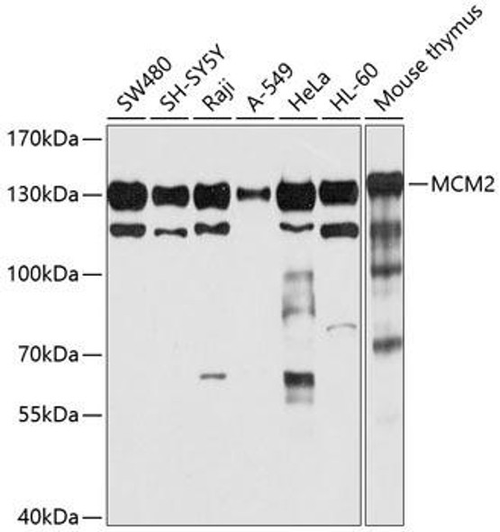
| Calculated MW: | 101kDa |
| Observed MW: | 125KDa |
| Synonyms: | MCM2, BM28, CCNL1, CDCL1, D3S3194, DFNA70, MITOTIN, cdc19 |
| Background: | The protein encoded by this gene is one of the highly conserved mini-chromosome maintenance proteins (MCM) that are involved in the initiation of eukaryotic genome replication. The hexameric protein complex formed by MCM proteins is a key component of the pre-replication complex (pre_RC) and may be involved in the formation of replication forks and in the recruitment of other DNA replication related proteins. This protein forms a complex with MCM4, 6, and 7, and has been shown to regulate the helicase activity of the complex. This protein is phosphorylated, and thus regulated by, protein kinases CDC2 and CDC7. Multiple alternatively spliced transcript variants have been found, but the full-length nature of some variants has not been defined. |
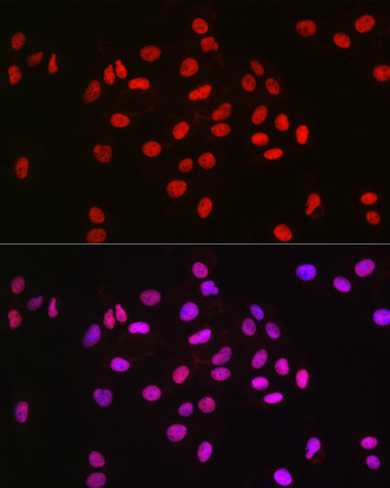 | Immunofluorescence analysis of U2OS cells using MCM2 Rabbit mAb at dilution of 1:100 (40x lens). Blue: DAPI for nuclear staining. |
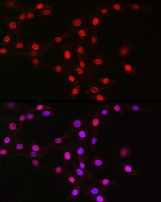 | Immunofluorescence analysis of NIH/3T3 cells using MCM2 Rabbit mAb at dilution of 1:100 (40x lens). Blue: DAPI for nuclear staining. |
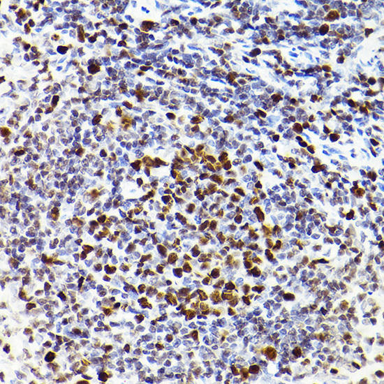 | Immunohistochemistry of paraffin-embedded rat spleen using MCM2 Rabbit mAb at dilution of 1:100 (40x lens). Perform high pressure antigen retrieval with 10 mM citrate buffer pH 6. 0 before commencing with IHC staining protocol. |
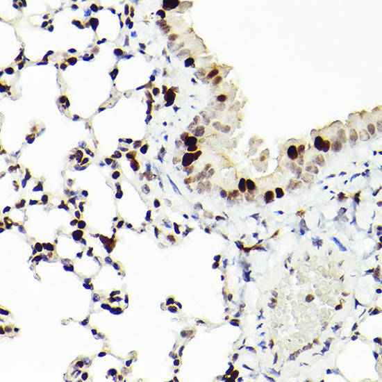 | Immunohistochemistry of paraffin-embedded mouse lung using MCM2 Rabbit mAb at dilution of 1:100 (40x lens). Perform high pressure antigen retrieval with 10 mM citrate buffer pH 6. 0 before commencing with IHC staining protocol. |
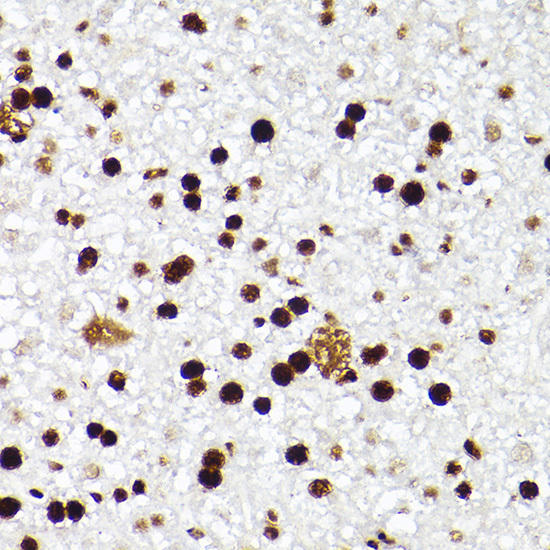 | Immunohistochemistry of paraffin-embedded mouse liver using MCM2 Rabbit mAb at dilution of 1:100 (40x lens). Perform high pressure antigen retrieval with 10 mM citrate buffer pH 6. 0 before commencing with IHC staining protocol. |
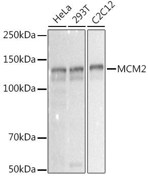 | Western blot analysis of extracts of various cell lines, using MCM2 antibody at 1:1000 dilution. Secondary antibody: HRP Goat Anti-Rabbit IgG (H+L) at 1:10000 dilution. Lysates/proteins: 25ug per lane. Blocking buffer: 3% nonfat dry milk in TBST. Detection: ECL Basic Kit. Exposure time: 0. 3s. |


