
Anti-Malondialdehyde Antibody [11E3]
- SKU:
- STMA00185
- Antibody Type:
- Monoclonal
- Applications:
- WB
- Applications:
- ICC/IF
- Applications:
- ELISA
- Applications:
- IHC
- Reactivity:
- Species Independent
- Host Species:
- Mouse
- Isotype:
- IgG1




Description
Malondialdehyde Antibody [11E3]
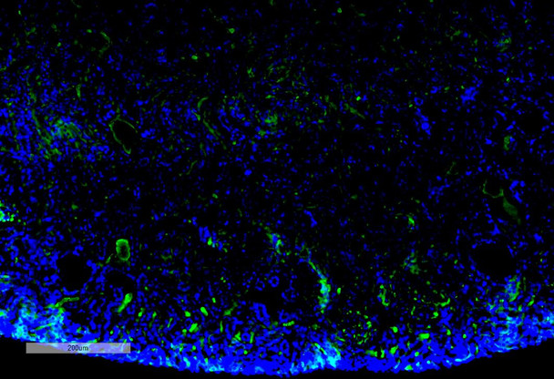
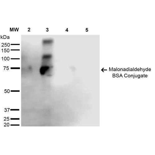
Malondialdehyde Antibody [11E3] is a mouse monoclonal antibody directed against Malondialdehyde, offered by Assay Genie for research applications. Reported applications include WB, ICC/IF, ELISA and IHC. Reactivity is species independent. Supplied as IgG1, purified by Protein G.
Malondialdehyde (MDA) is a highly reactive, low-molecular-weight aldehyde and one of the most widely used biomarkers of oxidative stress due to its chemical stability and ease of detection. Formed as a byproduct of lipid peroxidation, MDA reflects oxidative damage to polyunsaturated fatty acids in cellular membranes—a process central to the pathology of neurodegenerative diseases. MDA exhibits a strong affinity for amino groups, forming adducts with free amino acids, proteins, and nucleic acids. These adducts can disrupt protein function and induce DNA cross-linking, contributing to mutagenesis and cellular dysfunction. Elevated MDA levels have been consistently observed in Alzheimer’s disease and other neurodegenerative conditions, as well as in systemic diseases such as diabetes and cancer. In neuroscience research, MDA serves not only as a diagnostic and prognostic marker but also as a mechanistic link between oxidative stress and neuronal injury. Its quantification—commonly performed using the thiobarbituric acid reactive substances (TBARS) assay—provides valuable insights into the redox state of the brain and the efficacy of antioxidant therapies. Given its dual role as both a biomarker and a mediator of oxidative damage, MDA is a critical target in neurodegenerative disease research. Understanding and mitigating MDA-associated toxicity may offer new avenues for therapeutic intervention in oxidative stress-driven neurological disorders. This antibody is also available conjugated to ATTO 390, ATTO 488, ATTO 594, APC, Biotin, FITC, HRP, PerCP and RPE. Supplied in 100 µg. For research use only; not for diagnostic or therapeutic procedures.
| Product Name: | Malondialdehyde Antibody [11E3] |
| Product SKU: | STMA00185 |
| Size: | 100 µg |
| Target: | Malondialdehyde |
| Host Species: | Mouse |
| Antibody Type: | Monoclonal |
| Clone ID: | 11E3 |
| Research Areas: | Cancer, Oxidative Stress, Lipid peroxidation, Neuroscience, Neurodegeneration, Alzheimer's Disease |
| Buffer: | PBS pH7.4, 50% glycerol, 0.09% sodium azide |
| Storage & Shipping: | Store at -20°C. Shipped on Blue Ice or 4°C. Conjugated variants should be stored according to the product label. |
| Format: | Liquid |
| Applications: | WB, ICC/IF, ELISA, IHC |
| Antibody Isotype: | IgG1 |
| Purification: | Protein G Purified |
| Concentration: | 1 mg/ml |
| Reactivity: | Species Independent |
| Recommended Dilution: | WB (1:1000); ICC/IF (1:50); ELISA (1:1000); IHC (1:100); optimal dilutions for assays should be determined by the user. |
| Specificity: | Specific for Malondialdehyde conjugated proteins. Does not detect free Malondialdehyde. Does not cross-react with Acrolein, Crotonaldehyde, Hexanoyl Lysine, 4-Hydroxy-2-hexenal, 4-Hydroxy nonenal, or Methylglyoxal modified proteins. |
| Guarantee: | 12 months from date of dispatch |
| Immunogen: | Synthetic Malondialdehyde modified Keyhole Limpet Hemocyanin (KLH). |
| Alternative Names: | Malondialdehyde, MDA, Malondialdehyde (MDA), Malonic aldehyde, Propanedial, 1,3-Propanedial, Malonaldehyde |
| Note: | For research use only. |
Related Products

Anti-Malondialdehyde Antibody [6H6]

MDA (Malondialdehyde) ELISA Kit (UNES00040)
Hormone & Small Molecule ELISA Kits

Chicken Malondialdehyde (MDA) ELISA Kit (CHEB0536)
Chicken Immunology ELISA Kits

