Anti-HSP70/HSC70 Antibody [BB70]

(No reviews yet) Write a Review
SKU:
STMA00004
Antibody Type:
Monoclonal
Applications:
WB
Applications:
ICC/IF
Applications:
IHC
Applications:
IP
Reactivity:
Human
Reactivity:
Mouse
Reactivity:
Rat
Reactivity:
Bovine
Reactivity:
Sheep
Reactivity:
Dog
Reactivity:
Beluga
Reactivity:
Fish
Reactivity:
Guinea Pig (Cavia porcellus)
Reactivity:
Pig
Reactivity:
Hamster
Reactivity:
Rabbit
Reactivity:
Chicken
Reactivity:
African clawed frog (Xenopus laevis)
Reactivity:
Fruit Fly (Drosophila melanogaster)
Reactivity:
Yeast
Host Species:
Mouse
Isotype:
IgG2a
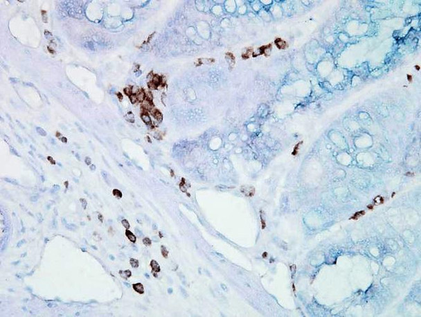
  • Immunohistochemistry analysis using Mouse Anti-Hsp70 Monoclonal Antibody, Clone BB70 (STMA00004). Tissue: inflamed colon. Species: Mouse. Fixation: Formalin. Primary Antibody: Mouse Anti-Hsp70 Monoclonal Antibody (STMA00004) at 1:10000 for 12 hours at 4°C. Secondary Antibody: Biotin Goat Anti-Mouse at 1:2000 for 1 hour at RT. Counterstain: Mayer Hematoxylin (purple/blue) nuclear stain at 200 µl for 2 minutes at RT.
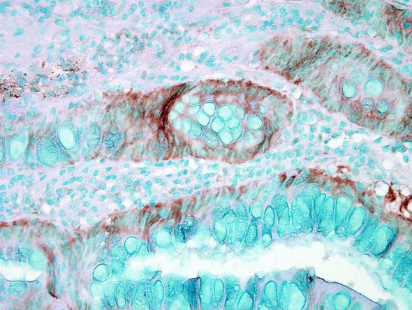
  • Immunohistochemistry analysis using Mouse Anti-Hsp70 Monoclonal Antibody, Clone BB70 (STMA00004). Tissue: colon carcinoma. Species: Human. Fixation: Formalin. Primary Antibody: Mouse Anti-Hsp70 Monoclonal Antibody (STMA00004) at 1:10000 for 12 hours at 4°C. Secondary Antibody: Biotin Goat Anti-Mouse at 1:2000 for 1 hour at RT. Counterstain: Mayer Hematoxylin (purple/blue) nuclear stain at 200 µl for 2 minutes at RT.
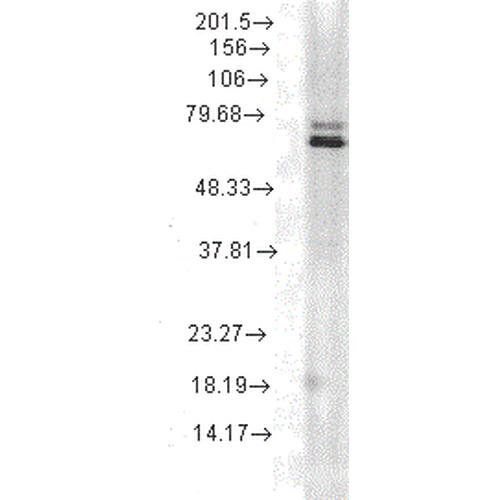
  • Western Blot analysis of Bovine MDBK cell lysates showing detection of Hsp70 protein using Mouse Anti-Hsp70 Monoclonal Antibody, Clone BB70 (STMA00004). Primary Antibody: Mouse Anti-Hsp70 Monoclonal Antibody (STMA00004) at 1:1000.
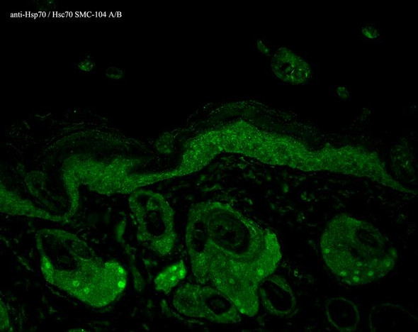
  • Immunocytochemistry/Immunofluorescence analysis using Mouse Anti-Hsp70 Monoclonal Antibody, Clone BB70 (STMA00004). Tissue: hepatocyte nuclei. Species: Rat. Primary Antibody: Mouse Anti-Hsp70 Monoclonal Antibody (STMA00004) at 1:200. Liver sections were paraffin embedded. First pictures in series show two hours after exposure to stress, the second shows the control. Courtesy of: G. Matic, University of Belgrade, Serbia.
  • Western Blot analysis of Human Cervical cancer cell line (HeLa) lysate showing detection of Hsp70 protein using Mouse Anti-Hsp70 Monoclonal Antibody, Clone BB70 (STMA00004). Primary Antibody: Mouse Anti-Hsp70 Monoclonal Antibody (STMA00004) at 1:1000. Secondary Antibody: HRP Goat Anti-Rat.
  • Immunohistochemistry analysis using Mouse Anti-Hsp70 Monoclonal Antibody, Clone BB70 (STMA00004). Tissue: hepatocytes. Species: Rat. Fixation: Paraffin Embedded. Primary Antibody: Mouse Anti-Hsp70 Monoclonal Antibody (STMA00004) at 1:200. Liver sections were paraffin embedded. First pictures in series show two hours after exposure to stress, the second shows the control. Courtesy of: G. Matic, University of Belgrade, Serbia.
Frequently bought together:

Description

HSP70/HSC70 Antibody [BB70]

HSP70/HSC70 Antibody [BB70] is a mouse monoclonal antibody directed against HSP70/HSC70, offered by Assay Genie for research applications. Reported applications include WB, ICC/IF, IHC and IP. Reported reactivity: Human, Mouse, Rat, Bovine, Sheep, Dog, Beluga, Fish, Guinea Pig (Cavia porcellus), Pig, Hamster, Rabbit, Chicken, African clawed frog (Xenopus laevis), Fruit Fly (Drosophila melanogaster) and Yeast. Supplied as IgG2a, purified by Protein G.

HSP70 proteins are a highly conserved family of 70-kDa molecular chaperones essential for maintaining protein homeostasis across all domains of life. In eukaryotes, HSP70 genes form a multigene family, with isoforms localized to the cytosol, nucleus, mitochondria, endoplasmic reticulum, and chloroplasts. These chaperones are either constitutively expressed (HSC70) or stress-inducible (HSP70), and share over 50% sequence identity across species. Functionally, HSP70s bind ATP with high affinity and exhibit ATPase activity that is stimulated by interaction with unfolded or misfolded proteins. The N-terminal domain mediates ATP binding, while the C-terminal domain is responsible for substrate recognition. This ATP-driven cycle enables HSP70s to bind and release hydrophobic regions of nascent or damaged proteins, preventing aggregation and facilitating proper folding, transport, and assembly. In neuroscience, HSP70 and HSC70 are critical for neuronal survival under proteotoxic stress—conditions commonly associated with neurodegenerative diseases such as Alzheimer’s, Parkinson’s, and Huntington’s disease. These chaperones stabilize misfolded proteins, support autophagic clearance, and modulate apoptotic signaling pathways. Their ability to interact with co-chaperones and degradation machinery further enhances their role in maintaining neuronal proteostasis. Given their central role in stress response and protein quality control, HSP70/HSC70 are promising therapeutic targets and biomarkers in neurodegeneration research. This antibody is also available conjugated to ATTO 390, ATTO 488, ATTO 594, APC, Biotin, FITC, HRP, PerCP and RPE. Supplied in 200 µg. For research use only; not for diagnostic or therapeutic procedures.

View AllClose

0 Reviews

View AllClose