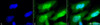
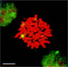
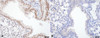
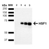
HSF1 Antibody [10H8] is a rat monoclonal antibody directed against HSF1, offered by Assay Genie for research applications. Reported applications include WB, IHC, ICC/IF, IP, ELISA and GS. Reported reactivity: Human, Mouse, Rat, Bovine, Guinea Pig (Cavia porcellus), Hamster, Monkey and Rabbit. Supplied as IgG1, purified by Protein G.
Heat Shock Factor 1 (HSF1) is a key transcriptional regulator that orchestrates the cellular response to proteotoxic stress by activating genes involved in protein folding, degradation, and quality control. As the primary mediator of the heat shock response, HSF1 binds to heat shock elements (HSEs) in the promoters of heat shock protein (HSP) genes, driving the expression of molecular chaperones essential for maintaining proteostasis. Structurally, HSF1 contains a conserved N-terminal DNA-binding domain and adjacent hydrophobic repeats critical for trimerization and activation. A second hydrophobic region near the C-terminus suppresses trimer formation under non-stress conditions. In unstressed cells, HSF1 is diffusely localized in the cytoplasm and nucleus. Upon exposure to stressors—such as oxidative damage, heat, or protein misfolding—HSF1 rapidly translocates to nuclear stress granules, initiating a transcriptional program that restores protein homeostasis. In the context of neurodegenerative diseases, HSF1 plays a pivotal role in mitigating the toxic accumulation of misfolded proteins, a hallmark of disorders such as Alzheimer’s, Parkinson’s, and Huntington’s disease. By enhancing the expression of protective HSPs, HSF1 supports neuronal survival and function under chronic stress conditions. Its dynamic regulation and neuroprotective potential make HSF1 a compelling therapeutic target in neuroscience and neurodegeneration research. This antibody is also available conjugated to ATTO 390, ATTO 488, ATTO 594, APC, Biotin, FITC, HRP, PerCP and RPE. Supplied in 100 µg. For research use only; not for diagnostic or therapeutic procedures.
1. Cotto J.J., Fox S.G. and Morimoto R.I. (1997) J. Cell Science 110: 2925-2934. 2. Morano K.A. and Thiele D.J. (1999). Gene Expression 7 (6): 271-82. 3.Tanaka KI et al. (2007). JBC Papers Online Manuscript M704081200. 4. Morimoto R. I. (1998) Genes Dev 12: 3788-3796. 5. McMillan D. R., Xiao X., Shao L., Graves K., and Benjamin I. J. (1998) J Bio Chem 273: 7523-7528. 6. Jolly C., Usson Y. and Morimoto R.I. (1999) Proc. Natl. Acad. Sci. USA 96 (12): 6769- 6774. 7. Fiorenza M.T., Farkas T., Dissing M., Kolding D. and Zimarino V. (1995) Nucleic Acids Res. 23 (3):467-474. 8. Goodson M.L., Park-Sarge O.K. and Sarge K.D. (1995) Mol. Cell. Biol. 15(10): 5288-5293. 9. Rallu M., et al. (1997) Proc. Natl. Acad. Sci. USA 94(6): 2392-2397. 10. Sarge K.D., et al. (1994) Biol. Reprod. 50(6): 1334- 1343. 11. Murphy S.P., Gorzowski J.J., Sarge K.D. and Phillips B. (1994) Mol. Cell. Biol. 14(8):5309-5317.
HSF1 Antibody [10H4]HSF1 Antibody [10H4] is a rat monoclonal antibody directed against HSF1, offered by Assay Genie for research applications. Reported applications include WB, IHC, ICC/IF, IP, ELISA...
HSF1 Antibody [4B4]HSF1 Antibody [4B4] is a rat monoclonal antibody directed against HSF1, offered by Assay Genie for research applications. Reported applications include WB, IHC, ICC/IF, IP, ELISA...






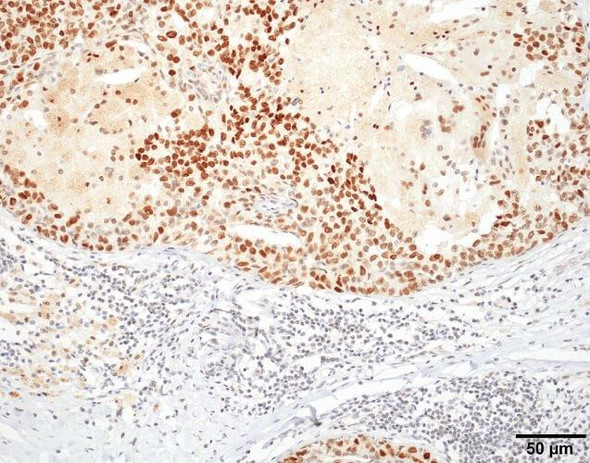


![Anti-EpCAM [1G8-8C7-10H8] Monoclonal Antibody (AGMB06104)](https://cdn11.bigcommerce.com/s-rd6ounxcu2/images/stencil/590x590/products/118043/132084/anti-epcam-1g8-8c7-10h8-monoclonal-antibody-agmb06104__83320__28190.1786851716.jpg?c=1 "Anti-EpCAM [1G8-8C7-10H8] Monoclonal Antibody (AGMB06104)")
![Anti-HSF1 [R09-3M8] Monoclonal Antibody (AGMB01030)](https://cdn11.bigcommerce.com/s-rd6ounxcu2/images/stencil/590x590/products/112979/127022/anti-hsf1-r09-3m8-monoclonal-antibody-agmb01030__55865__89220.1786838724.jpg?c=1 "Anti-HSF1 [R09-3M8] Monoclonal Antibody (AGMB01030)")
![Anti- HSF1 [R07-0G0] Monoclonal Antibody - Knockout Validated (AGMB06590)](https://cdn11.bigcommerce.com/s-rd6ounxcu2/images/stencil/590x590/products/118529/132569/anti-hsf1-r07-0g0-monoclonal-antibody-knockout-validated-agmb06590__05665__30533.1786852959.jpg?c=1 "Anti- HSF1 [R07-0G0] Monoclonal Antibody - Knockout Validated (AGMB06590)")