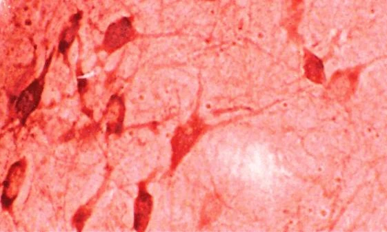
HO-1 Antibody [1F12-A6] is a mouse monoclonal antibody directed against HO-1, offered by Assay Genie for research applications. Reported applications include WB, IHC, ICC/IF, IP and ELISA. Reported reactivity: Human, Mouse, Rat, Bovine, Dog, Guinea Pig (Cavia porcellus), Hamster, Monkey, Pig and Rabbit. Supplied as IgG1 Kappa, purified by Protein G.
Heme oxygenase-1 (HO-1), also known as heat shock protein 32, is an inducible enzyme that catalyzes the degradation of heme into biliverdin, free iron, and carbon monoxide—each with distinct biological functions. Biliverdin is rapidly converted to bilirubin, a potent antioxidant, while carbon monoxide acts as a signaling molecule with anti-inflammatory and vasodilatory properties. Free iron, although potentially toxic, is sequestered by ferritin to limit oxidative damage. HO-1 is strongly upregulated in response to oxidative stress, inflammation, and neurotoxic insults, positioning it as a key cytoprotective factor in the central nervous system. Its expression is elevated in various neurodegenerative conditions, including Alzheimer’s disease, Parkinson’s disease, and multiple sclerosis, where it helps mitigate neuronal injury by reducing reactive oxygen species and modulating immune responses. Unlike its constitutive isoform HO-2, HO-1 is dynamically regulated and serves as a frontline defense against cellular stress. Deficiency in HO-1 has been linked to increased neuroinflammation, impaired stress tolerance, and heightened vulnerability to neurodegenerative pathology. Given its dual role in detoxifying free heme and generating neuroprotective metabolites, HO-1 is a promising therapeutic target for modulating oxidative stress and inflammation in neurodegenerative disease research. This antibody is also available conjugated to ATTO 390, ATTO 488, ATTO 594, APC, Biotin, FITC, HRP, PerCP and RPE. Supplied in 100 µg. For research use only; not for diagnostic or therapeutic procedures.
1. Froh M. et al. (2007) World J. Gastroentereol 13(25): 3478-86. 2. Elbirt K.K. and Bonkovsky H.L. (1999) Proc Assoc Am Physicians 111(5): 348-47. 3. Maines M.D., Trakshel G.M., and Kutty R.K. (1986) J Biol Chem 261: 411–419. 4. Brydun A., et al. (2007) Hypertens Res 30(4): 341-8. 5. Poss K.D. and Tonegawa S. (1997). Proc Natl Acad Sci U S A. 94: 10925–10930. 6. Yet S.F., et al. (2003) FASEB J. 17: 1759–1761. 7. Yachie A., et al. (1999) J Clin Invest. 103: 129–135.
HO-1 AntibodyHO-1 Antibody is a rabbit polyclonal antibody directed against HO-1, offered by Assay Genie for research applications. Reported applications include WB, IHC and IP. Reported reactivity:...
HO-1 AntibodyHO-1 Antibody is a rabbit polyclonal antibody directed against HO-1, offered by Assay Genie for research applications. Reported applications include WB, ICC/IF and IHC. Reported...
HO-1 Antibody [6B8-2F2]HO-1 Antibody [6B8-2F2] is a mouse monoclonal antibody directed against HO-1, offered by Assay Genie for research applications. Reported applications include WB and ICC/IF...



![Anti-cIAP2 (1F12) [1F12-E5-G12] Monoclonal Antibody (AGMB04238)](https://cdn11.bigcommerce.com/s-rd6ounxcu2/images/stencil/590x590/products/116188/130225/anti-ciap2-1f12-1f12-e5-g12-monoclonal-antibody-agmb04238__10548__56336.1786846956.jpg?c=1 "Anti-cIAP2 (1F12) [1F12-E5-G12] Monoclonal Antibody (AGMB04238)")
![Anti-Eg5 (4H3) [4H3-1F12] Monoclonal Antibody (AGMB04290)](https://cdn11.bigcommerce.com/s-rd6ounxcu2/images/stencil/590x590/products/116238/130274/anti-eg5-4h3-4h3-1f12-monoclonal-antibody-agmb04290__68665__80171.1786847084.jpg?c=1 "Anti-Eg5 (4H3) [4H3-1F12] Monoclonal Antibody (AGMB04290)")