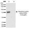
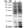
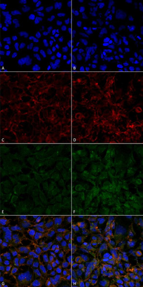
Hexanoyl-Lysine adduct Antibody [5D9] is a mouse monoclonal antibody directed against Hexanoyl-Lysine adduct, offered by Assay Genie for research applications. Reported applications include WB, ICC/IF, FACS, FCM, ELISA and IHC. Reactivity is species independent. Supplied as IgG1, purified by Protein G.
Hexanoyl-lysine adduct (HEL) is a lipid peroxidation marker formed by the reaction of oxidized fatty acids with lysine residues on proteins. It serves as an early indicator of oxidative stress, a central feature in the pathogenesis of neurodegenerative diseases. In the brain, HEL accumulation reflects damage to neuronal membranes and proteins caused by reactive oxygen species (ROS). Elevated levels of HEL have been detected in models of Alzheimer’s disease, Parkinson’s disease, and other neurodegenerative conditions, correlating with disease progression and cognitive decline. HEL is particularly valuable as a biomarker due to its stability and specificity for lipid peroxidation. Its presence indicates oxidative damage at an early stage, often preceding overt neurodegeneration. This makes HEL a promising candidate for early diagnosis, disease monitoring, and evaluating the efficacy of antioxidant therapies. By highlighting the oxidative component of neurodegeneration, HEL contributes to a deeper understanding of disease mechanisms and supports the development of targeted interventions aimed at reducing oxidative injury in the nervous system. This antibody is also available conjugated to ATTO 390, ATTO 488, ATTO 594, APC, Biotin, FITC, HRP, PerCP and RPE. Supplied in 100 µg. For research use only; not for diagnostic or therapeutic procedures.
Product Name:
Hexanoyl-Lysine adduct Antibody [5D9]
Product SKU:
STMA00179
Size:
100 µg
Target:
Hexanoyl-Lysine adduct
Host Species:
Mouse
Antibody Type:
Monoclonal
Clone ID:
5D9
Research Areas:
Cancer, Oxidative Stress, Lipid peroxidation
Buffer:
PBS pH7.4, 50% glycerol, 0.09% sodium azide
Storage & Shipping:
Store at -20°C. Shipped on Blue Ice or 4°C. Conjugated variants should be stored according to the product label.
Format:
Liquid
Applications:
WB, ICC/IF, FACS, FCM, ELISA, IHC
Antibody Isotype:
IgG1
Purification:
Protein G Purified
Concentration:
1 mg/ml
Reactivity:
Species Independent
Recommended Dilution:
WB (1:1000); ICC/IF (1:50); ELISA (1:1000); FACS (1:50); FCM (1:50); IHC (1:100); optimal dilutions for assays should be determined by the user.
Specificity:
Specific for Hexanoyl-Lysine adduct (HEL) modified peptides and proteins. Does not detect free Hexanoyl-Lysine. Does not cross-react with Acrolein, Crotonaldehyde, 4-Hydroxy-2-hexenal, 4-Hydroxy nonenal, Malondialdehyde, or Methylglyoxal modified proteins.
Hexanoyl-Lysine adduct Antibody [5E8]Hexanoyl-Lysine adduct Antibody [5E8] is a mouse monoclonal antibody directed against Hexanoyl-Lysine adduct, offered by Assay Genie for research applications...
Acetylated Lysine AntibodyAcetylated Lysine Antibody is a rabbit polyclonal antibody directed against Acetylated Lysine, offered by Assay Genie for research applications. Reported applications...
Methylated Lysine AntibodyMethylated Lysine Antibody is a rabbit polyclonal antibody directed against Methylated Lysine, offered by Assay Genie for research applications. Reported applications...





![Anti-CaMKII alpha (5D9) [5D9-G10-G8] Monoclonal Antibody (AGMB04615)](https://cdn11.bigcommerce.com/s-rd6ounxcu2/images/stencil/590x590/products/116562/130605/anti-camkii-alpha-5d9-5d9-g10-g8-monoclonal-antibody-agmb04615__51339__16283.1786847930.jpg?c=1 "Anti-CaMKII alpha (5D9) [5D9-G10-G8] Monoclonal Antibody (AGMB04615)")



![Anti-EAAT1 [R05-5D9] Monoclonal Antibody (AGMB01410)](https://cdn11.bigcommerce.com/s-rd6ounxcu2/images/stencil/590x590/products/113360/127404/anti-eaat1-r05-5d9-monoclonal-antibody-agmb01410__73920__06680.1786839702.jpg?c=1 "Anti-EAAT1 [R05-5D9] Monoclonal Antibody (AGMB01410)")