Description
| Antibody Name: | Anti-GSDMD Antibody (CAB20728) |
| Antibody SKU: | CAB20728 |
| Antibody Size: | 50µL, 100µL |
| Application: | Western blotting, Immunofluorescence |
| Reactivity: | Human, Mouse, Rat |
| Host Species: | Rabbit |
| Immunogen: | Recombinant fusion protein containing a sequence corresponding to amino acids 346-484 of human GSDMD (NP_079012.3). |
| Application: | Western blotting, Immunofluorescence |
| Recommended Dilution: | WB 1:500 - 1:2000 IF 1:50 - 1:200 |
| Reactivity: | Human, Mouse, Rat |
| Positive Samples: | THP-1, A-431 |
| Immunogen: | Recombinant fusion protein containing a sequence corresponding to amino acids 346-484 of human GSDMD (NP_079012.3). |
| Purification Method: | Affinity purification |
| Storage Buffer: | Store at -20°C. Avoid freeze / thaw cycles. Buffer: PBS with 0.02% sodium azide, 50% glycerol, pH7.3. |
| Isotype: | IgG |
| Sequence: | DGPA GAVL ECLV LSSG MLVP ELAI PVVY LLGA LTML SETQ HKLL AEAL ESQT LLGP LELV GSLL EQSA PWQE RSTM SLPP GLLG NSWG EGAP AWVL LDEC GLEL GEDT PHVC WEPQ AQGR MCAL YASL ALLS GLSQ EPH |
| Cellular Location: | |
| Calculated MW: | 53kDa |
| Observed MW: | 53KDa |
| Synonyms: | DF5L, DFNA5L, FKSG10, GSDMDC1, GSDMD |
| Background: | Gasdermin D is a member of the gasdermin family. Members of this family appear to play a role in regulation of epithelial proliferation. Gasdermin D has been suggested to act as a tumor suppressor. Alternatively spliced transcript variants have been described. |
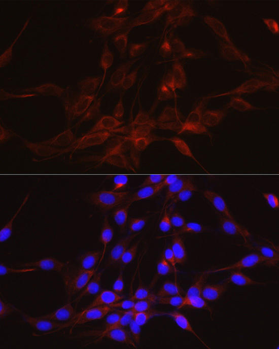 | Immunofluorescence analysis of PC-12 cells using GSDMD Rabbit mAb at dilution of 1:100 (40x lens). Blue: DAPI for nuclear staining. |
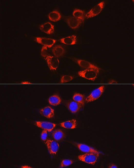 | Immunofluorescence analysis of NIH/3T3 cells using GSDMD Rabbit mAb at dilution of 1:100 (40x lens). Blue: DAPI for nuclear staining. |
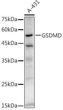 | Western blot analysis of extracts of A-431 cells, using GSDMD antibody at 1:1000 dilution. Secondary antibody: HRP Goat Anti-Rabbit IgG (H+L) at 1:10000 dilution. Lysates/proteins: 25ug per lane. Blocking buffer: 3% nonfat dry milk in TBST. Detection: ECL Enhanced Kit. Exposure time: 180s. |
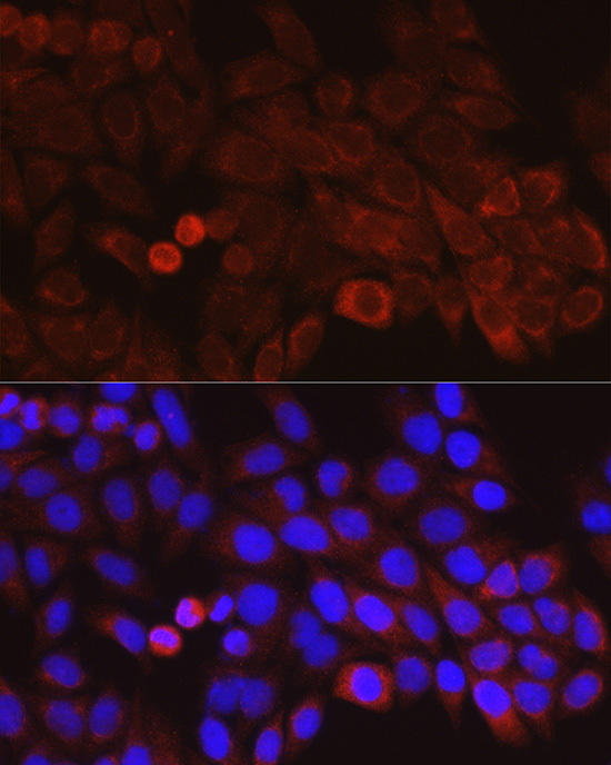 | Immunofluorescence analysis of HeLa cells using GSDMD Rabbit mAb at dilution of 1:100 (40x lens). Blue: DAPI for nuclear staining. |
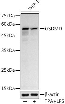 | Western blot analysis of extracts of THP-1 cells, using GSDMD antibody at 1:1000 dilution. THP-1 cells were treated by LPS (1 μg/ml) at 37℃ for 8 hours. THP-1 cells were treated by PMA/TPA (200 nM) at 37℃ for 15 minutes after serum-starvation overnight. Secondary antibody: HRP Goat Anti-Rabbit IgG (H+L) at 1:10000 dilution. Lysates/proteins: 25ug per lane. Blocking buffer: 3% nonfat dry milk in TBST. Detection: ECL Basic Kit. Exposure time: 180s. |


