GFP Antibody [H24] is a mouse recombinant monoclonal antibody directed against GFP, offered by Assay Genie for research applications. Reported applications include WB and ICC/IF. Reactivity is species independent. Supplied as IgG1, purified by Protein A.
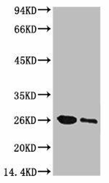
Green fluorescent protein (GFP) is a 27 kDa reporter protein originally isolated from the jellyfish Aequorea victoria. It emits bright green fluorescence when exposed to ultraviolet or blue light, making it an indispensable tool in molecular and cellular neuroscience. GFP enables real-time visualization of gene expression, protein localization, and cellular dynamics in live cells and tissues without the need for additional substrates or cofactors. In neurodegenerative disease research, GFP is widely used to track neuronal morphology, monitor synaptic changes, and study the progression of protein aggregation in models of Alzheimer’s, Parkinson’s, and Huntington’s disease. By fusing GFP to disease-relevant proteins such as tau, α-synuclein, or huntingtin, researchers can observe pathological processes in real time and assess the efficacy of therapeutic interventions. The recombinant nature of GFP ensures high specificity, consistent expression, and minimal cytotoxicity, which enhances reproducibility across experimental systems. Additionally, its compatibility with advanced imaging techniques—including confocal and two-photon microscopy—makes GFP a cornerstone in the study of neural circuits and neurodegenerative mechanisms. As a genetically encoded fluorescent marker, GFP continues to drive innovation in neuroscience by enabling precise, dynamic, and non-invasive investigation of cellular processes underlying brain function and disease. This antibody is also available conjugated to ATTO 390, ATTO 488, ATTO 594, APC, Biotin, FITC, HRP, PerCP and RPE. Supplied in 100 µg. For research use only; not for diagnostic or therapeutic procedures.
Product Name:
GFP Antibody [H24]
Product SKU:
STMA00231
Size:
100 µg
Target:
GFP
Host Species:
Mouse
Antibody Type:
Recombinant Monoclonal
Clone ID:
H24
Research Areas:
Tags and Cell Markers, Fluorescent Proteins
Buffer:
PBS pH 7.4, 50% glycerol, 0.09% NaN3
Storage & Shipping:
Store at -20°C. Shipped on Blue Ice or 4°C. Conjugated variants should be stored according to the product label.
Format:
Liquid
Applications:
WB, ICC/IF
Antibody Isotype:
IgG1
Purification:
Protein A Purified
Concentration:
1mg/ml
Reactivity:
Species Independent
Recommended Dilution:
WB (1:5000), ICC/IF (1:100); optimal dilutions for assays should be determined by the user.
Specificity:
Detects full length GFP
Guarantee:
12 months from date of dispatch
Immunogen:
Fusion protein, AA #1-238 (full length) of jellyfish GFP
Immunogen Species:
Jellyfish
Swiss-Prot:
P42212
Alternative Names:
GFP, Green fluorescent protein
References:
1. Prasher, D. C., Eckenrode, V. K., Ward, W. W., Prendergast, F. G., & Cormier, M. J. (1992). Primary structure of the Aequorea victoria green-fluorescent protein. Gene, 111(2), 229–233. DOI: 10.1016/0378-1119(92)90691-H 2. Tsien, R. Y. (1998). The green fluorescent protein. Annual Review of Biochemistry, 67(1), 509–544. DOI: 10.1146/annurev.biochem.67.1.509 3. Bradbury, A., & Plückthun, A. (2015). Reproducibility: Standardize antibodies used in research. Nature, 518(7537), 27–29. DOI: 10.1038/518027a

![Western blot analysis with Mouse Anti-GFP Monoclonal Antibody, Clone H24 (STMA00231) showing detection of recombinant Aequorea GFP protein, his-tagged (Invitrogen A42613). Block: 2% skim milk + 2% BSA for 30 minutes at RT. Primary Antibody: Mouse Anti-Jellyfish GFP Recombinant Monoclonal [H24] (STMA00231) at 1:5000 for 1 hour at RT. Secondary Antibody: Goat anti-mouse IgG-HRP (H&L) at 1:5000 for 30 minutes at RT.](https://cdn11.bigcommerce.com/s-rd6ounxcu2/images/stencil/1280x1280/products/120578/135670/anti-gfp-antibody-h24__71211__30641.1786858495.jpg?c=1?imbypass=on "Western blot analysis with Mouse Anti-GFP Monoclonal Antibody, Clone H24 (STMA00231) showing detection of recombinant Aequorea GFP protein, his-tagged (Invitrogen A42613). Block: 2% skim milk + 2% BSA for 30 minutes at RT. Primary Antibody: Mouse Anti-Jellyfish GFP Recombinant Monoclonal [H24] (STMA00231) at 1:5000 for 1 hour at RT. Secondary Antibody: Goat anti-mouse IgG-HRP (H&L) at 1:5000 for 30 minutes at RT.")

![Western blot analysis with Mouse Anti-GFP Monoclonal Antibody, Clone H24 (STMA00231) showing detection of recombinant Aequorea GFP protein, his-tagged (Invitrogen A42613). Block: 2% skim milk + 2% BSA for 30 minutes at RT. Primary Antibody: Mouse Anti-Jellyfish GFP Recombinant Monoclonal [H24] (STMA00231) at 1:5000 for 1 hour at RT. Secondary Antibody: Goat anti-mouse IgG-HRP (H&L) at 1:5000 for 30 minutes at RT.](https://cdn11.bigcommerce.com/s-rd6ounxcu2/images/stencil/608x608/products/120578/135670/anti-gfp-antibody-h24__71211__30641.1786858495.jpg?c=1 "Western blot analysis with Mouse Anti-GFP Monoclonal Antibody, Clone H24 (STMA00231) showing detection of recombinant Aequorea GFP protein, his-tagged (Invitrogen A42613). Block: 2% skim milk + 2% BSA for 30 minutes at RT. Primary Antibody: Mouse Anti-Jellyfish GFP Recombinant Monoclonal [H24] (STMA00231) at 1:5000 for 1 hour at RT. Secondary Antibody: Goat anti-mouse IgG-HRP (H&L) at 1:5000 for 30 minutes at RT.")

![Anti-GFP Tag [R04-6A1] Monoclonal Antibody (AGMB06832)](https://cdn11.bigcommerce.com/s-rd6ounxcu2/images/stencil/590x590/products/118773/132810/anti-gfp-tag-r04-6a1-monoclonal-antibody-agmb06832__04521__88702.1786853578.jpg?c=1 "Anti-GFP Tag [R04-6A1] Monoclonal Antibody (AGMB06832)")