Anti-ERp19 Antibody (CAB9386)
")
- SKU:
- CAB9386
- Product Type:
- Antibody
- Antibody Type:
- Monoclonal Antibody
- Reactivity:
- Human
- Reactivity:
- Mouse
- Reactivity:
- Rat
- Host Species:
- Rabbit
- Isotype:
- IgG
")
Description
| Antibody Name: | Anti-ERp19 Antibody (CAB9386) |
| Antibody SKU: | CAB9386 |
| Antibody Size: | 50µL, 100µL |
| Application: | Western blotting, Immunohistochemistry |
| Reactivity: | Human, Mouse, Rat |
| Host Species: | Rabbit |
| Immunogen: | A synthesized peptide derived from human ERp19. |
| Application: | Western blotting, Immunohistochemistry |
| Recommended Dilution: | WB 1:500 - 1:2000 IHC 1:50 - 1:200 |
| Reactivity: | Human, Mouse, Rat |
| Positive Samples: | HT-29, 293T, SH-SY5Y, Mouse lung, Mouse heart, Mouse placenta, Rat lung, Rat kidney, Rat heart |
| Immunogen: | A synthesized peptide derived from human ERp19. |
| Purification Method: | Affinity purification |
| Storage Buffer: | Store at -20°C. Avoid freeze / thaw cycles. Buffer: PBS with 0.02% sodium azide, 50% glycerol, pH7.3. |
| Isotype: | IgG |
| Sequence: | Email for sequence |
| Cellular Location: | Endoplasmic reticulum lumen |
| Calculated MW: | 17kDa |
| Observed MW: | 19KDa |
| Synonyms: | TXNDC12, AG1, AGR1, ERP16, ERP18, ERP19, PDIA16, TLP19, hAG-1, hTLP19, thioredoxin domain containing 12 |
| Background: | This gene encodes a member of the thioredoxin superfamily. Members of this family are characterized by a conserved active motif called the thioredoxin fold that catalyzes disulfide bond formation and isomerization. This protein localizes to the endoplasmic reticulum and has a single atypical active motif. The encoded protein is mainly involved in catalyzing native disulfide bond formation and displays activity similar to protein-disulfide isomerases. This protein may play a role in defense against endoplasmic reticulum stress. Alternate splicing results in both coding and non-coding variants. |
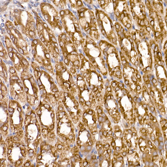 | Immunohistochemistry of paraffin-embedded mouse stomach using ERp19 Rabbit mAb at dilution of 1:200 (40x lens). Perform high pressure antigen retrieval with 10 mM citrate buffer pH 6. 0 before commencing with IHC staining protocol. |
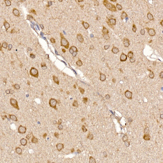 | Immunohistochemistry of paraffin-embedded rat brain using ERp19 Rabbit mAb at dilution of 1:200 (40x lens). Perform high pressure antigen retrieval with 10 mM citrate buffer pH 6. 0 before commencing with IHC staining protocol. |
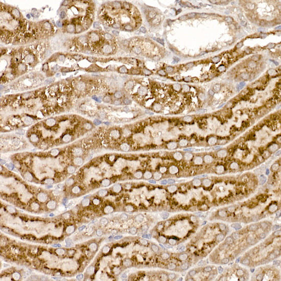 | Immunohistochemistry of paraffin-embedded mouse kidney using ERp19 Rabbit mAb at dilution of 1:200 (40x lens). Perform high pressure antigen retrieval with 10 mM citrate buffer pH 6. 0 before commencing with IHC staining protocol. |
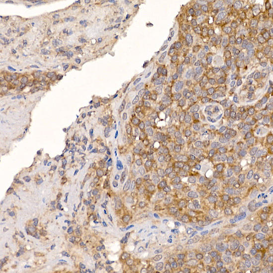 | Immunohistochemistry of paraffin-embedded human tonsil using ERp19 Rabbit mAb at dilution of 1:200 (40x lens). Perform high pressure antigen retrieval with 10 mM citrate buffer pH 6. 0 before commencing with IHC staining protocol. |
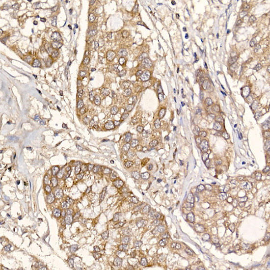 | Immunohistochemistry of paraffin-embedded human lung cancer using ERp19 Rabbit mAb at dilution of 1:200 (40x lens). Perform high pressure antigen retrieval with 10 mM citrate buffer pH 6. 0 before commencing with IHC staining protocol. |
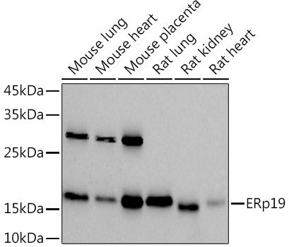 | Western blot analysis of extracts of various cell lines, using at 1:1000 dilution. Secondary antibody: HRP Goat Anti-Rabbit IgG (H+L) at 1:10000 dilution. Lysates/proteins: 25ug per lane. Blocking buffer: 3% nonfat dry milk in TBST. Detection: ECL Basic Kit. Exposure time: 90s. |
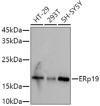 | Western blot analysis of extracts of various cell lines, using at 1:1000 dilution. Secondary antibody: HRP Goat Anti-Rabbit IgG (H+L) at 1:10000 dilution. Lysates/proteins: 25ug per lane. Blocking buffer: 3% nonfat dry milk in TBST. Detection: ECL Basic Kit. Exposure time: 3s. |
Related Products

 ELISA Kit")
Human anti-EPO antibody(anti-Erythropoietin antibody) ELISA Kit
system_update_altDatasheet - तकनीकी मैनुअलsystem_update_altMSDS - तकनीक
")
 ELISA Kit")
Human Anti-DSG3 antibody(anti-Desmoglein-3 antibody) ELISA Kit
system_update_altDatasheet - तकनीकी मैनुअलsystem_update_altMSDS - तकनीक
")
Anti-YUCCA1 Antibody (CAB20733)
OverviewAntibody Name:Anti-YUCCA1 Antibody (CAB20733)Antibody SKU:CAB20733Antibody Size:50µL, 100µLApplication:Western blottingReactivi