Description
| Antibody Name: | Anti-Cystatin B Antibody (CAB20886) |
| Antibody SKU: | CAB20886 |
| Antibody Size: | 50µL, 100µL |
| Application: | Western blotting |
| Reactivity: | Human, Mouse, Rat |
| Host Species: | Rabbit |
| Immunogen: | Recombinant protein of Human Cystatin B. |
| Application: | Western blotting |
| Recommended Dilution: | WB 1:500 - 1:2000 |
| Reactivity: | Human, Mouse, Rat |
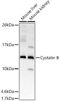
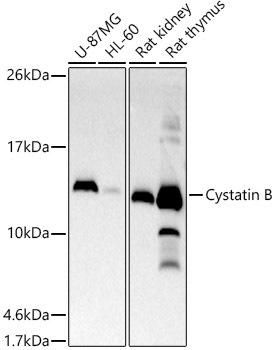
| Positive Samples: | U-87MG, HL-60, Mouse liver, Mouse kidney, Rat kidney, Rat thymus |
| Immunogen: | Recombinant protein of Human Cystatin B. |
| Purification Method: | Affinity purification |
| Storage Buffer: | Store at -20°C. Avoid freeze / thaw cycles. Buffer: PBS with 0.02% sodium azide, 0.05% BSA, 50% glycerol, pH7.3. |
| Isotype: | IgG |
| Sequence: | Email for sequence |
| Cellular Location: | Cytoplasm, Nucleus |
| Calculated MW: | 11kDa |
| Observed MW: | 13KDa |
| Synonyms: | CSTB, CPI-B, CST6, EPM1, EPM1A, PME, STFB, ULD |
| Background: | The cystatin superfamily encompasses proteins that contain multiple cystatin-like sequences. Some of the members are active cysteine protease inhibitors, while others have lost or perhaps never acquired this inhibitory activity. There are three inhibitory families in the superfamily, including the type 1 cystatins (stefins), type 2 cystatins and kininogens. This gene encodes a stefin that functions as an intracellular thiol protease inhibitor. The protein is able to form a dimer stabilized by noncovalent forces, inhibiting papain and cathepsins l, h and b. The protein is thought to play a role in protecting against the proteases leaking from lysosomes. Evidence indicates that mutations in this gene are responsible for the primary defects in patients with progressive myoclonic epilepsy (EPM1). One type of mutation responsible for EPM1 is the expansion in the promoter region of this gene of a CCCCGCCCCGCG repeat from 2-3 copies to 30-78 copies. |
 | Western blot analysis of extracts of various cell lines, using Cystatin B antibody at 1:500 dilution. Secondary antibody: HRP Goat Anti-Rabbit IgG (H+L) at 1:10000 dilution. Lysates/proteins: 25ug per lane. Blocking buffer: 3% nonfat dry milk in TBST. Detection: ECL Basic Kit. Exposure time: 180s. |
 | Western blot analysis of extracts of various cell lines, using Cystatin B antibody at 1:500 dilution. Secondary antibody: HRP Goat Anti-Rabbit IgG (H+L) at 1:10000 dilution. Lysates/proteins: 25ug per lane. Blocking buffer: 3% nonfat dry milk in TBST. Detection: ECL Basic Kit. Exposure time: 30s. |


