Description
| Antibody Name: | Anti-CD68 Antibody (CAB20803) |
| Antibody SKU: | CAB20803 |
| Antibody Size: | 50µL, 100µL |
| Application: | Western blotting, Immunohistochemistry |
| Reactivity: | Human, Mouse, Rat |
| Host Species: | Rabbit |
| Immunogen: | A synthetic peptide corresponding to a sequence within amino acids 100-200 of human CD68 (NP_001242.2). |
| Application: | Western blotting, Immunohistochemistry |
| Recommended Dilution: | WB 1:500 - 1:2000 IHC 1:50 - 1:200 |
| Reactivity: | Human, Mouse, Rat |
| Positive Samples: | Raji, THP-1, U-937, RAW264.7, Mouse spleen, Rat lung |
| Immunogen: | A synthetic peptide corresponding to a sequence within amino acids 100-200 of human CD68 (NP_001242.2). |
| Purification Method: | Affinity purification |
| Storage Buffer: | Store at -20°C. Avoid freeze / thaw cycles. Buffer: PBS with 0.02% sodium azide, 50% glycerol, pH7.3. |
| Isotype: | IgG |
| Sequence: | TSQG PSTA THSP ATTS HGNA TVHP TSNS TATS PGFT SSAH PEPP PPSP SPSP TSKE TIGD YTWT NGSQ PCVH LQAQ IQIR VMYT TQGG GEAW GISV LNPN K |
| Cellular Location: | Cell membrane, Endosome membrane, Lysosome membrane, Single-pass type I membrane protein, Single-pass type I membrane protein |
| Calculated MW: | 31kDa/34kDa/37kDa |
| Observed MW: | 80-130KDa |
| Synonyms: | CD68, GP110, LAMP4, SCARD1 |
| Background: | This gene encodes a 110-kD transmembrane glycoprotein that is highly expressed by human monocytes and tissue macrophages. It is a member of the lysosomal/endosomal-associated membrane glycoprotein (LAMP) family. The protein primarily localizes to lysosomes and endosomes with a smaller fraction circulating to the cell surface. It is a type I integral membrane protein with a heavily glycosylated extracellular domain and binds to tissue- and organ-specific lectins or selectins. The protein is also a member of the scavenger receptor family. Scavenger receptors typically function to clear cellular debris, promote phagocytosis, and mediate the recruitment and activation of macrophages. Alternative splicing results in multiple transcripts encoding different isoforms. |
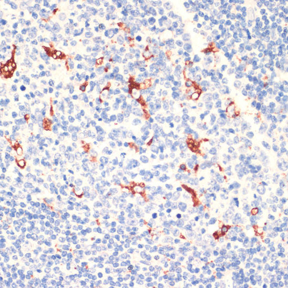
 | Immunohistochemistry of paraffin-embedded human tonsil using CD68 Rabbit mAb at dilution of 1:72900 (40x lens). Perform high pressure antigen retrieval with 10 mM citrate buffer pH 6. 0 before commencing with IHC staining protocol. |
 | Immunohistochemistry of paraffin-embedded human colon using CD68 Rabbit mAb at dilution of 1:8100 (40x lens). Perform high pressure antigen retrieval with 10 mM citrate buffer pH 6. 0 before commencing with IHC staining protocol. |
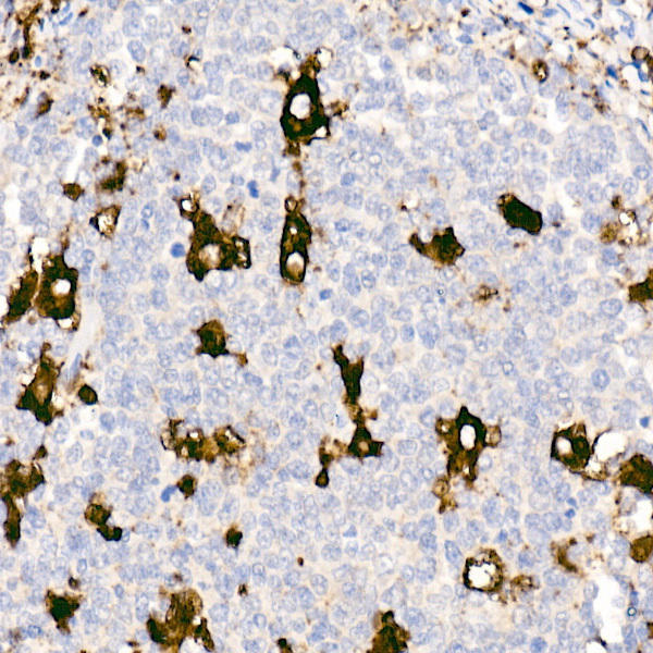
 | Immunohistochemistry of paraffin-embedded human tonsil using CD68 antibody at dilution of 1:50 (40x lens). Perform high pressure antigen retrieval with 10 mM citrate buffer pH 6. 0 before commencing with IHC staining protocol. |
 | Immunohistochemistry of paraffin-embedded human colon using CD68 Rabbit mAb at dilution of 1:72900 (40x lens). Perform high pressure antigen retrieval with 10 mM citrate buffer pH 6. 0 before commencing with IHC staining protocol. |
 | Immunohistochemistry of paraffin-embedded human colon using CD68 antibody at dilution of 1:50 (40x lens). Perform high pressure antigen retrieval with 10 mM citrate buffer pH 6. 0 before commencing with IHC staining protocol. |
 | Immunohistochemistry of paraffin-embedded human brain using CD68 antibody at dilution of 1:50 (40x lens). Perform high pressure antigen retrieval with 10 mM citrate buffer pH 6. 0 before commencing with IHC staining protocol. |
 | Immunohistochemistry of paraffin-embedded human lung using CD68 antibody at dilution of 1:50 (40x lens). Perform high pressure antigen retrieval with 10 mM citrate buffer pH 6. 0 before commencing with IHC staining protocol. |
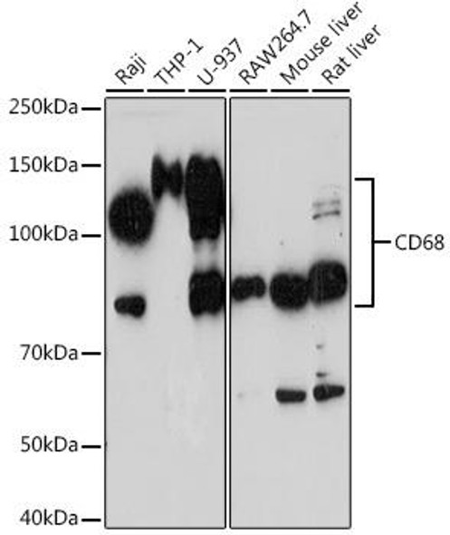
 | Western blot analysis of extracts of various cell lines, using CD68 antibody at 1:1000 dilution. Secondary antibody: HRP Goat Anti-Rabbit IgG (H+L) at 1:10000 dilution. Lysates/proteins: 25ug per lane. Blocking buffer: 3% nonfat dry milk in TBST. Detection: ECL Basic Kit. Exposure time: 90s. |
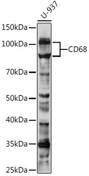
 | Western blot analysis of extracts of various cell lines, using CD68 antibody at 1:1000 dilution. Secondary antibody: HRP Goat Anti-Rabbit IgG (H+L) at 1:10000 dilution. Lysates/proteins: 25ug per lane. Blocking buffer: 3% nonfat dry milk in TBST. Detection: ECL Basic Kit. Exposure time: 30s. |


