Anti-AK2 Antibody [KO Validated] (CAB20934)
![Anti-AK2 Antibody [KO Validated] (CAB20934)](https://cdn11.bigcommerce.com/s-rd6ounxcu2/images/stencil/608x608/products/78128/83310/anti-ak2-antibody-ko-validated-cab20934__46275__59053.1706545086.jpg?c=1 "Anti-AK2 Antibody [KO Validated] (CAB20934)")
- SKU:
- CAB20934
![Anti-AK2 Antibody [KO Validated] (CAB20934)](https://cdn11.bigcommerce.com/s-rd6ounxcu2/images/stencil/1280x1280/products/78128/83310/anti-ak2-antibody-ko-validated-cab20934__46275__59053.1706545086.jpg?c=1?imbypass=on "Anti-AK2 Antibody [KO Validated] (CAB20934)")
Description
| Antibody Name: | Anti-AK2 Antibody [KO Validated] (CAB20934) |
| Antibody SKU: | CAB20934 |
| Antibody Size: | 50µL, 100µL |
| Application: | Western blotting, Immunofluorescence |
| Reactivity: | Human, Mouse, Rat |
| Host Species: | Rabbit |
| Immunogen: | Recombinant protein of Human AK2. |
| Application: | Western blotting, Immunofluorescence |
| Recommended Dilution: | WB 1:500 - 1:2000 IF 1:50 - 1:200 |
| Reactivity: | Human, Mouse, Rat |
| Positive Samples: | HeLa, HepG2, Mouse liver, Mouse kidney, Mouse heart, Rat liver, Rat kidney |
| Immunogen: | Recombinant protein of Human AK2. |
| Purification Method: | Affinity purification |
| Storage Buffer: | Store at -20°C. Avoid freeze / thaw cycles. Buffer: PBS with 0.02% sodium azide, 0.05% BSA, 50% glycerol, pH7.3. |
| Isotype: | IgG |
| Sequence: | Email for sequence |
| Cellular Location: | Mitochondrion intermembrane space |
| Calculated MW: | 14kDa/21kDa/22kDa/24kDa/25kDa/26kDa |
| Observed MW: | 26KDa |
| Synonyms: | AK2, ADK2 |
| Background: | Adenylate kinases are involved in regulating the adenine nucleotide composition within a cell by catalyzing the reversible transfer of phosphate groups among adenine nucleotides. Three isozymes of adenylate kinase, namely 1, 2, and 3, have been identified in vertebrates; this gene encodes isozyme 2. Expression of these isozymes is tissue-specific and developmentally regulated. Isozyme 2 is localized in the mitochondrial intermembrane space and may play a role in apoptosis. Mutations in this gene are the cause of reticular dysgenesis. Alternate splicing results in multiple transcript variants. Pseudogenes of this gene are found on chromosomes 1 and 2. |
![Immunofluorescence analysis of NIH/3T3 cells using [KO Validated] AK2 Rabbit mAb at dilution of 1:25 (40x lens). Blue: DAPI for nuclear staining.](https://cdn11.bigcommerce.com/s-h68l9z2lnx/product_images/m/030/anti-ak2-antibody-ko-validated-cab20934__46275.jpg) | Immunofluorescence analysis of NIH/3T3 cells using [KO Validated] AK2 Rabbit mAb at dilution of 1:25 (40x lens). Blue: DAPI for nuclear staining. |
![Immunofluorescence analysis of MCF7 cells using [KO Validated] AK2 Rabbit mAb at dilution of 1:25 (40x lens). Blue: DAPI for nuclear staining.](https://cdn11.bigcommerce.com/s-h68l9z2lnx/product_images/j/402/anti-ak2-antibody-ko-validated-cab20934__74068.jpg) | Immunofluorescence analysis of MCF7 cells using [KO Validated] AK2 Rabbit mAb at dilution of 1:25 (40x lens). Blue: DAPI for nuclear staining. |
![Immunofluorescence analysis of HepG2 cells using [KO Validated] AK2 Rabbit mAb at dilution of 1:25 (40x lens). Blue: DAPI for nuclear staining.](https://cdn11.bigcommerce.com/s-h68l9z2lnx/product_images/i/371/anti-ak2-antibody-ko-validated-cab20934__40234.jpg) | Immunofluorescence analysis of HepG2 cells using [KO Validated] AK2 Rabbit mAb at dilution of 1:25 (40x lens). Blue: DAPI for nuclear staining. |
![Immunofluorescence analysis of HeLa cells using [KO Validated] AK2 Rabbit mAb at dilution of 1:25 (40x lens). Blue: DAPI for nuclear staining.](https://cdn11.bigcommerce.com/s-h68l9z2lnx/product_images/n/731/anti-ak2-antibody-ko-validated-cab20934__25895.jpg) | Immunofluorescence analysis of HeLa cells using [KO Validated] AK2 Rabbit mAb at dilution of 1:25 (40x lens). Blue: DAPI for nuclear staining. |
 | Western blot analysis of extracts from normal (control) and AK2 knockout (KO) HeLa cells, using AK2 antibody at 1:500 dilution. Secondary antibody: HRP Goat Anti-Rabbit IgG (H+L) at 1:10000 dilution. Lysates/proteins: 25ug per lane. Blocking buffer: 3% nonfat dry milk in TBST. Detection: ECL Basic Kit. Exposure time: 1s. |
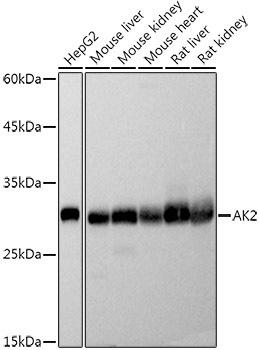 | Western blot analysis of extracts of various cell lines, using AK2 antibody at 1:500 dilution. Secondary antibody: HRP Goat Anti-Rabbit IgG (H+L) at 1:10000 dilution. Lysates/proteins: 25ug per lane. Blocking buffer: 3% nonfat dry milk in TBST. Detection: ECL Basic Kit. Exposure time: 1s. |
Additional Information
Product Type: |
Antibody |
Antibody Type: |
Polyclonal Antibody |
Reactivity: |
Human |
Reactivity: |
Mouse |
Reactivity: |
Rat |
Host Species: |
Rabbit |
Isotype: |
IgG |
Related Products
![Anti-AK2 Antibody (CAB6519)[KO Validated]](https://cdn11.bigcommerce.com/s-rd6ounxcu2/images/stencil/590x590/products/46800/51544/anti-ak2-antibody-cab6519ko-validated__07752__27828.1706525077.jpg?c=1 "Anti-AK2 Antibody (CAB6519)[KO Validated]")
![Anti-CASP9 Antibody [KO Validated]](https://cdn11.bigcommerce.com/s-rd6ounxcu2/images/stencil/590x590/products/55751/60933/anti-casp9-antibody-ko-validated__51716__83049.1706533665.jpg?c=1 "Anti-CASP9 Antibody [KO Validated]")
![Anti-STAT3 Antibody (CAB1192)[KO Validated]](https://cdn11.bigcommerce.com/s-rd6ounxcu2/images/stencil/590x590/products/39393/44139/anti-stat3-antibody-cab1192ko-validated__14333__14709.1706521315.jpg?c=1 "Anti-STAT3 Antibody (CAB1192)[KO Validated]")
![Anti-MLKL Antibody (CAB13451)[KO Validated]](https://cdn11.bigcommerce.com/s-rd6ounxcu2/images/stencil/590x590/products/40763/45508/anti-mlkl-antibody-cab13451ko-validated__79871__53808.1706521997.jpg?c=1 "Anti-MLKL Antibody (CAB13451)[KO Validated]")
![Anti-MMP9 Antibody (CAB2095)[KO Validated]](https://cdn11.bigcommerce.com/s-rd6ounxcu2/images/stencil/590x590/products/44713/49458/anti-mmp9-antibody-cab2095ko-validated__86648__78803.1706524016.jpg?c=1 "Anti-MMP9 Antibody (CAB2095)[KO Validated]")