Anti-Acetyl-Histone H3-K23 Antibody (CAB2771)
- SKU:
- CAB2771
- Product Type:
- Antibody
- Antibody Type:
- Monoclonal Antibody
- Applications:
- WB
- Applications:
- IHC
- Applications:
- IF
- Applications:
- IP
- Applications:
- ChIP
- Reactivity:
- Human
- Reactivity:
- Mouse
- Reactivity:
- Rat
- Host Species:
- Rabbit
- Isotype:
- IgG
- Research Area:
- Epigenetics and Nuclear Signaling
Description
| Product Name: | Acetyl-Histone H3-K23 Rabbit mAb |
| Product Code: | CAB2771 |
| Size: | 20uL, 50uL, 100uL |
| Applications: | WB, IHC, IF, IP, ChIP |
| Reactivity: | Human, Mouse, Rat |
| Host Species: | Rabbit |
| Immunogen: | A synthesized peptide derived from human Histone H3 (acetyl K27). |
| Applications: | WB, IHC, IF, IP, ChIP |
| Recommended Dilutions: | WB 1:500 - 1:2000 IHC 1:50 - 1:200 IF 1:50 - 1:200 ChIP 1:50 - 1:200 |
| Reactivity: | Human, Mouse, Rat |
| Positive Samples: | HeLa, NIH/3T3, C6 |
| Immunogen: | A synthesized peptide derived from human Histone H3 (acetyl K27). |
| Purification Method: | Affinity purification |
| Storage: | Store at -20°C. Avoid freeze / thaw cycles. Buffer: PBS with 0.02% sodium azide, 50% glycerol, pH7.3. |
| Isotype: | IgG |
| Sequence: | Email for sequence |
| Gene ID: | 8350/3020/8290/440093/126961 |
| Uniprot: | P68431/P84243/Q16695/Q6NXT2/Q71DI3 |
| Calculated MW: | 17 kDa |
| Observed MW: | 17KDa |
| UniProt Protein Function: | H3: Core component of nucleosome. Nucleosomes wrap and compact DNA into chromatin, limiting DNA accessibility to the cellular machineries which require DNA as a template. Histones thereby play a central role in transcription regulation, DNA repair, DNA replication and chromosomal stability. DNA accessibility is regulated via a complex set of post-translational modifications of histones, also called histone code, and nucleosome remodeling. The nucleosome is a histone octamer containing two molecules each of H2A, H2B, H3 and H4 assembled in one H3-H4 heterotetramer and two H2A-H2B heterodimers. The octamer wraps approximately 147 bp of DNA. Belongs to the histone H3 family. |
| UniProt Protein Details: | Protein type:DNA-binding Chromosomal Location of Human Ortholog: 6p22.2 Cellular Component: extracellular region; membrane; nuclear chromosome; nuclear chromosome, telomeric region; nucleoplasm; nucleosome; nucleus; protein complex Molecular Function:cadherin binding; histone binding; protein binding Biological Process: blood coagulation; cellular protein metabolic process; chromatin silencing at rDNA; DNA replication-dependent nucleosome assembly; establishment and/or maintenance of chromatin architecture; negative regulation of gene expression, epigenetic; nucleosome assembly; positive regulation of gene expression, epigenetic; protein heterotetramerization; RNA-mediated gene silencing; telomere organization and biogenesis |
| NCBI Summary: | Histones are basic nuclear proteins that are responsible for the nucleosome structure of the chromosomal fiber in eukaryotes. Two molecules of each of the four core histones (H2A, H2B, H3, and H4) form an octamer, around which approximately 146 bp of DNA is wrapped in repeating units, called nucleosomes. The linker histone, H1, interacts with linker DNA between nucleosomes and functions in the compaction of chromatin into higher order structures. This gene is intronless and encodes a replication-dependent histone that is a member of the histone H3 family. Transcripts from this gene lack polyA tails but instead contain a palindromic termination element. This gene is found in the small histone gene cluster on chromosome 6p22-p21.3. [provided by RefSeq, Aug 2015] |
| UniProt Code: | P68431 |
| NCBI GenInfo Identifier: | 55977055 |
| NCBI Gene ID: | 8357 |
| NCBI Accession: | P68431.2 |
| UniProt Secondary Accession: | P68431,P02295, P02296, P16106, Q6ISV8, Q6NWP8, Q6NWP9 Q6NXU4, Q71DJ3, Q93081, A0PJT7, A5PLR1, |
| UniProt Related Accession: | P68431 |
| Molecular Weight: | 15kDa |
| NCBI Full Name: | Histone H3.1 |
| NCBI Synonym Full Names: | histone cluster 1 H3 family member h |
| NCBI Official Symbol: | HIST1H3H |
| NCBI Official Synonym Symbols: | H3/k; H3FK; H3F1K |
| NCBI Protein Information: | histone H3.1 |
| UniProt Protein Name: | Histone H3.1 |
| UniProt Synonym Protein Names: | Histone H3/a; Histone H3/b; Histone H3/c; Histone H3/d; Histone H3/f; Histone H3/h; Histone H3/i; Histone H3/j; Histone H3/k; Histone H3/l |
| UniProt Gene Name: | HIST1H3A |
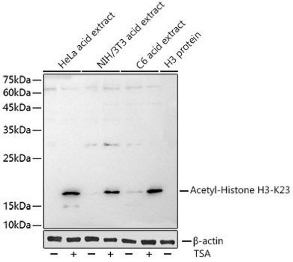
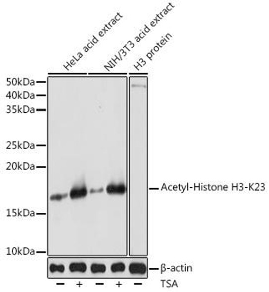
 | Western blot analysis of extracts of various cell lines, using Acetyl-Histone H3-K27 antibody at 1:1000 dilution. HeLa NIH/3T3 and C6 cells were treated by TSA (1 uM) at 37°C for 18 hours. Secondary antibody: HRP Goat Anti-Rabbit IgG (H+L) at 1:10000 dilution. Lysates/proteins: 25ug per lane. Blocking buffer: 3% nonfat dry milk in TBST. Detection: ECL Basic Kit. Exposure time: 180s. |
 | Immunohistochemistry of paraffin-embedded human breast cancer using Acetyl-Histone H3-K27 Rabbit mAb at dilution of 1:100 (40x lens). Perform high pressure antigen retrieval with 10 mM citrate buffer pH 6. 0 before commencing with IHC staining protocol. |
 | Immunohistochemistry of paraffin-embedded mouse lung using Acetyl-Histone H3-K27 Rabbit mAb at dilution of 1:100 (40x lens). Perform high pressure antigen retrieval with 10 mM citrate buffer pH 6. 0 before commencing with IHC staining protocol. |
 | Immunohistochemistry of paraffin-embedded rat kidney using Acetyl-Histone H3-K27 Rabbit mAb at dilution of 1:100 (40x lens). Perform high pressure antigen retrieval with 10 mM citrate buffer pH 6. 0 before commencing with IHC staining protocol. |
 | Immunofluorescence analysis of NIH/3T3 cells using Acetyl-Histone H3-K27 Rabbit mAb at dilution of 1:100 (40x lens). Blue: DAPI for nuclear staining. |
 | Immunofluorescence analysis of PC-12 cells using Acetyl-Histone H3-K27 Rabbit mAb at dilution of 1:100 (40x lens). Blue: DAPI for nuclear staining. |
 | Immunofluorescence analysis of U2OS cells using Acetyl-Histone H3-K27 Rabbit mAb at dilution of 1:100 (40x lens). Blue: DAPI for nuclear staining. |
 | Chromatin immunoprecipitation analysis of extracts of HeLa cells, using Acetyl-Histone H3-K27 antibody and rabbit IgG. The amount of immunoprecipitated DNA was checked by quantitative PCR. Histogram was constructed by the ratios of the immunoprecipitated DNA to the input. |


